

 No Comments
When your retina specialist tells you that you need a vitrectomy, the first reaction most patients have is fear – not because the word sounds serious (though it does), but because nobody has explained what it actually means. What is being removed? Will it hurt? Will you be awake? Will your vision come back?
These are the questions I hear every week in my clinic at Medanta Lucknow. And they deserve honest, detailed answers – not medical jargon that leaves you more confused than when you walked in.
In this post, I want to explain vitrectomy surgery from the ground up: what it is, why it is done, exactly what happens during the procedure, and what your recovery will realistically look like. If you or someone you love has been advised to have this surgery, read this first.
What Is Vitrectomy Surgery?
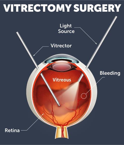
The word “vitrectomy” comes from vitreous – the clear, gel-like substance that fills the inside of your eye – and ectomy, meaning surgical removal. So at its most basic level, a vitrectomy is a procedure in which the vitreous gel inside your eye is carefully removed and replaced.
But why would anyone need to remove something that is supposed to be there?
The vitreous humor is mostly water and collagen. In a healthy young eye, it sits firmly against the retina. As we age – or due to disease, injury, or diabetes – this gel can bleed into itself, develop scar tissue, pull on the retina, or cloud over. When that happens, it stops being helpful and starts causing damage. Removing it gives the surgeon direct access to the retina to repair whatever is wrong, and the eye adapts remarkably well to its absence.
When Is Vitrectomy Needed? – Conditions That Require This Surgery
Vitrectomy is not a single-purpose procedure. It is used to treat a range of serious retinal and vitreous conditions, including:
Retinal Detachment: When the retina peels away from the back of the eye, a vitrectomy allows the surgeon to gently peel scar tissue, flatten the retina, and seal it back in place.
Vitreous Hemorrhage: Bleeding inside the eye – most commonly from diabetic retinopathy – clouds the vitreous with blood, blocking vision. Vitrectomy removes the blood-filled gel and restores clarity.
Diabetic Tractional Retinal Detachment: In advanced diabetic eye disease, scar tissue contracts and pulls the retina. Vitrectomy removes this scar tissue before permanent damage occurs.
Macular Hole: A hole that develops in the central retina (macula) – affecting reading and fine detail vision – can be closed with vitrectomy combined with a gas bubble.
Epiretinal Membrane (Macular Pucker): A thin layer of scar tissue grows over the macula, wrinkling it and distorting vision. Vitrectomy allows the membrane to be peeled away with fine forceps.
Endophthalmitis: Severe infection inside the eye requires vitrectomy to remove infected material and deliver antibiotics directly.
Foreign Body Removal: Objects that have entered the eye through trauma can be safely removed via vitrectomy.
In each of these cases, no other approach gives the surgeon the access, visibility, and control that vitrectomy provides.
Step by Step: What Actually Happens During Vitrectomy Surgery
This is the part most patients are most anxious about. Let me walk you through it exactly as it happens.
Before Surgery
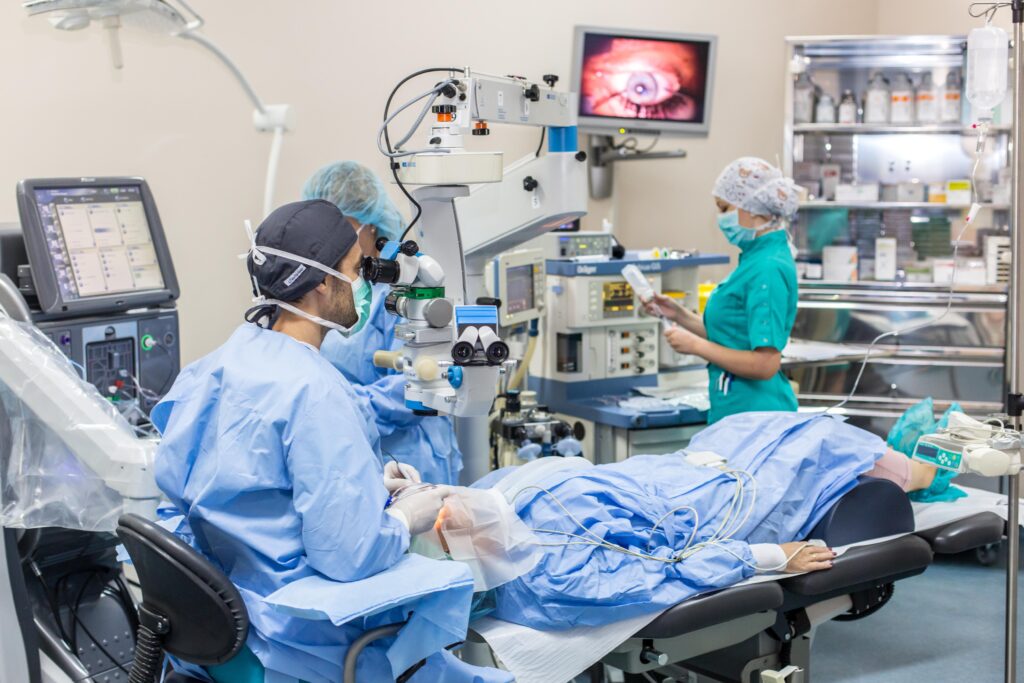
You will be asked to fast for a few hours before the procedure. Dilating eye drops will be applied to widen your pupil so the surgeon can see clearly. The area around your eye will be cleaned and sterile drapes placed. Most vitrectomies are performed under local anaesthesia with sedation – meaning your eye is completely numb, you are relaxed and comfortable, but you are not under general anaesthesia. You may be aware of light and some movement but you will feel nothing.
The Procedure – Three Small Incisions
Modern vitrectomy is performed through three tiny incisions (called ports) made in the white of the eye (sclera), each less than 1mm in size. Through these three ports, the surgeon introduces:
- A light pipe to illuminate the inside of the eye
- An infusion cannula to maintain eye pressure throughout surgery
- The vitrectomy cutter – a microsurgical instrument that simultaneously cuts and suctions the vitreous gel out of the eye at up to 10,000 cuts per minute
Working under a high-powered microscope, the surgeon removes the vitreous gel, then addresses the underlying condition – peeling membranes, sealing retinal tears with laser, flattening a detached retina, or removing scar tissue.
Tamponade – Holding the Retina in Place
Once the repair is done, the space left by the removed vitreous needs to be filled with something that holds the retina flat while it heals. The choice depends on the condition being treated:
- Gas bubble (SF6 or C3F8): Absorbs naturally over 2–8 weeks. During this time, patients must maintain a specific head position so the bubble stays against the repaired area.
- Silicone oil: Used in complex or high-risk cases. More durable than gas, but requires a second minor procedure to remove it once healing is complete, typically after 3–6 months.
- Balanced salt solution: Used in simpler cases where no tamponade is required.
The three tiny ports are then closed – often without stitches in modern small-gauge surgery – and a patch is placed over the eye.
Total surgical time: typically 1 to 3 hours, depending on the complexity of the underlying condition.
Will It Hurt?
During the surgery, no – the eye is fully anaesthetised. You may feel some pressure or movement, but pain is not expected. After surgery, mild aching or a gritty sensation is common for the first 24–48 hours and is managed with prescribed pain relief and anti-inflammatory drops.
Vitrectomy Recovery: A Realistic Week-by-Week Guide
Recovery from vitrectomy is where patient commitment matters most. What you do in the weeks after surgery significantly affects your outcome.
Day 1–3:
Your eye will be red, swollen, and your vision will be very blurry – particularly if a gas bubble was used. The bubble creates a dark, watery shadow in your visual field. This is completely normal. You will be given antibiotic and steroid eye drops to prevent infection and reduce inflammation. Rest is essential.
Days 4–7:
Swelling begins to settle. If a gas bubble is in place, you need to maintain the prescribed head position as directed – typically face-down or to one side. This is uncomfortable but critical. Missing position compliance is one of the most common reasons for incomplete retinal reattachment.
Weeks 2–4:
Most patients notice gradual improvement in vision as the gas bubble shrinks and the eye begins to clear. You may return to light activities and desk work, but no strenuous exercise, no bending below the waist, and no air travel until your surgeon confirms the gas has fully absorbed (air pressure changes can expand the gas and damage the eye).
Weeks 4–8:
Vision continues to stabilize. The rate of recovery depends on the original condition and how long it had been present before surgery. Macular hole closures, for example, can take 3–4 months to show full visual improvement.
3–6 months:
Final visual outcome becomes clearer. If silicone oil was used, your surgeon will plan its removal at this stage.
Important: Cataract formation is a very common side effect of vitrectomy in patients who still have their natural lens. Most patients will develop a visually significant cataract within 1–2 years of surgery. This is manageable with routine cataract surgery at the appropriate time.
What Results Can You Realistically Expect?
The outcome of vitrectomy depends heavily on three factors: the underlying condition, how long it had been present before surgery, and whether the macula (the central retina responsible for reading and detail) was involved.
In retinal detachment cases where the macula was still attached when surgery was performed, the chances of recovering useful central vision are very high. If the macula has already detached, recovery is possible but takes longer and may be incomplete.
For macular hole and epiretinal membrane cases, anatomical success (closing the hole or removing the membrane) is achieved in over 90% of cases. Functional visual improvement follows in the majority – but patience is required.
What I tell every patient before surgery: vitrectomy’s goal is first to save the eye, then to protect what vision remains, and then to improve it. In straightforward cases, all three are achievable. In advanced cases, the priority is preservation.
Why Choose a Vitreoretinal Specialist for This Surgery?
Vitrectomy is among the most technically demanding procedures in all of ophthalmology. It requires a surgeon trained specifically in vitreoretinal surgery – not a general eye surgeon. The difference matters enormously when complications arise, when membranes need peeling with instruments thinner than a human hair, or when a diabetic retina needs careful dissection.
At retinasurgeonlucknow.com, I have performed over 3,200 vitreoretinal surgeries, including complex diabetic tractional detachments, recurrent retinal detachments, and trauma cases. Every case is different, and the surgical plan is made specifically for each patient’s eye – not a one-size-fits-all approach.
Preparing for Your Vitrectomy: What to Do Before You Come In
If you have been advised to have vitrectomy surgery, here is how to prepare:
- Stop blood thinners (aspirin, warfarin, etc.) as advised by your doctor – usually 5–7 days before
- Arrange for someone to accompany you – you cannot drive after the procedure
- Arrange for help at home for the first week, particularly if face-down positioning is required
- Stop contact lenses a week before surgery
- Inform your surgeon of all medications, allergies, and any systemic conditions such as diabetes or hypertension
- Fast for 6–8 hours before surgery as instructed
A pre-surgery consultation with Dr. Charu Chaudhary will give you a personalised preparation plan and answer every question specific to your condition.
Frequently Asked Questions
Is vitrectomy a permanent fix?
In most cases, yes – the underlying repair is permanent. However, some conditions such as diabetic retinopathy can progress and affect the eye again over time, which is why ongoing monitoring is essential.
How soon can I go back to work?
Most patients with desk jobs return to work within 2–3 weeks. Physical or outdoor work takes 6–8 weeks. Your surgeon will advise based on your specific case.
Can vitrectomy be done in Lucknow?
Yes. Advanced vitreoretinal surgery, including micro-incision vitrectomy is performed at Medanta Lucknow. Patients no longer need to travel to Delhi or Mumbai for this procedure.
What if I need vitrectomy but I am diabetic?
Diabetic patients can undergo vitrectomy safely with proper blood sugar management before and after surgery. In fact, diabetic tractional retinal detachment is one of the most common reasons vitrectomy is performed.
Take the Next Step
If you have been told you need vitrectomy, or if you are experiencing symptoms – sudden floaters, flashes of light, a shadow across your vision, or unexplained vision loss – do not wait.
A consultation will determine the most appropriate treatment for your condition. The earlier we intervene, the better the outcome.
Book a Pre-Surgery Consultation →
Or learn more about retinal conditions we treat at our Retinal Surgery Service page.
Dr. Charu Chaudhary is a Vitreoretinal Surgeon at Medanta Lucknow with over 15 years of experience and 3,200+ surgical cases. She specialises in vitrectomy, retinal detachment repair, diabetic vitreoretinal surgery, and complex macular conditions. For appointments: Contact Us


Leave A Comment