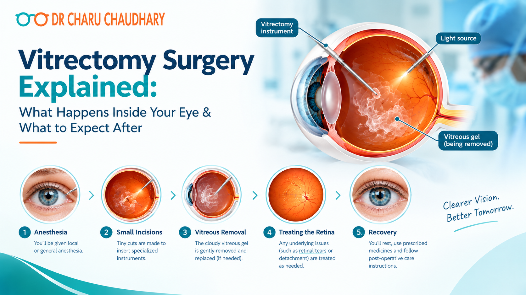
When your retina specialist tells you that you need a vitrectomy, the first reaction most patients have is fear – not because the word sounds serious (though it does), but because nobody has explained what it actually means. What is being removed? Will it hurt? Will you be awake? Will your vision come back? These are the questions I hear every week in my clinic at Medanta Lucknow. And they deserve honest, detailed answers – not medical jargon that leaves you more confused than when you walked in. In this post, I want to explain vitrectomy surgery from the ground up: what it is, why it is done, exactly what happens during the procedure, and what your recovery will realistically look like. If you or someone you love has been advised to have this surgery, read this first. What Is Vitrectomy Surgery? The word “vitrectomy” comes from vitreous – the clear, gel-like substance that fills the inside of your eye – and ectomy, meaning surgical removal. So at its most basic level, a vitrectomy is a procedure in which the vitreous gel inside your eye is carefully removed and replaced. But why would anyone need to remove something that is supposed to be there? The vitreous humor is mostly water and collagen. In a healthy young eye, it sits firmly against the retina. As we age – or due to disease, injury, or diabetes – this gel can bleed into itself, develop scar tissue, pull on the retina, or cloud over. When that happens, it stops being helpful and starts causing damage. Removing it gives the surgeon direct access to the retina to repair whatever is wrong, and the eye adapts remarkably well to its absence. When Is Vitrectomy Needed? – Conditions That Require This Surgery Vitrectomy is not a single-purpose procedure. It is used to treat a range of serious retinal and vitreous conditions, including: Retinal Detachment: When the retina peels away from the back of the eye, a vitrectomy allows the surgeon to gently peel scar tissue, flatten the retina, and seal it back in place. Vitreous Hemorrhage: Bleeding inside the eye – most commonly from diabetic retinopathy – clouds the vitreous with blood, blocking vision. Vitrectomy removes the blood-filled gel and restores clarity. Diabetic Tractional Retinal Detachment: In advanced diabetic eye disease, scar tissue contracts and pulls the retina. Vitrectomy removes this scar tissue before permanent damage occurs. Macular Hole: A hole that develops in the central retina (macula) – affecting reading and fine detail vision – can be closed with vitrectomy combined with a gas bubble. Epiretinal Membrane (Macular Pucker): A thin layer of scar tissue grows over the macula, wrinkling it and distorting vision. Vitrectomy allows the membrane to be peeled away with fine forceps. Endophthalmitis: Severe infection inside the eye requires vitrectomy to remove infected material and deliver antibiotics directly. Foreign Body Removal: Objects that have entered the eye through trauma can be safely removed via vitrectomy. In each of these cases, no other approach gives the surgeon the access, visibility, and control that vitrectomy provides. Step by Step: What Actually Happens During Vitrectomy Surgery This is the part most patients are most anxious about. Let me walk you through it exactly as it happens. Before Surgery You will be asked to fast for a few hours before the procedure. Dilating eye drops will be applied to widen your pupil so the surgeon can see clearly. The area around your eye will be cleaned and sterile drapes placed. Most vitrectomies are performed under local anaesthesia with sedation – meaning your eye is completely numb, you are relaxed and comfortable, but you are not under general anaesthesia. You may be aware of light and some movement but you will feel nothing. The Procedure – Three Small Incisions Modern vitrectomy is performed through three tiny incisions (called ports) made in the white of the eye (sclera), each less than 1mm in size. Through these three ports, the surgeon introduces: A light pipe to illuminate the inside of the eye An infusion cannula to maintain eye pressure throughout surgery The vitrectomy cutter – a microsurgical instrument that simultaneously cuts and suctions the vitreous gel out of the eye at up to 10,000 cuts per minute Working under a high-powered microscope, the surgeon removes the vitreous gel, then addresses the underlying condition – peeling membranes, sealing retinal tears with laser, flattening a detached retina, or removing scar tissue. Tamponade – Holding the Retina in Place Once the repair is done, the space left by the removed vitreous needs to be filled with something that holds the retina flat while it heals. The choice depends on the condition being treated: Gas bubble (SF6 or C3F8): Absorbs naturally over 2–8 weeks. During this time, patients must maintain a specific head position so the bubble stays against the repaired area. Silicone oil: Used in complex or high-risk cases. More durable than gas, but requires a second minor procedure to remove it once healing is complete, typically after 3–6 months. Balanced salt solution: Used in simpler cases where no tamponade is required. The three tiny ports are then closed – often without stitches in modern small-gauge surgery – and a patch is placed over the eye. Total surgical time: typically 1 to 3 hours, depending on the complexity of the underlying condition. Will It Hurt? During the surgery, no – the eye is fully anaesthetised. You may feel some pressure or movement, but pain is not expected. After surgery, mild aching or a gritty sensation is common for the first 24–48 hours and is managed with prescribed pain relief and anti-inflammatory drops. Vitrectomy Recovery: A Realistic Week-by-Week Guide Recovery from vitrectomy is where patient commitment matters most. What you do in the weeks after surgery significantly affects your outcome. Day 1–3: Your eye will be red, swollen, and your vision will be very blurry – particularly if a gas bubble was used. The bubble creates a dark, watery shadow in your
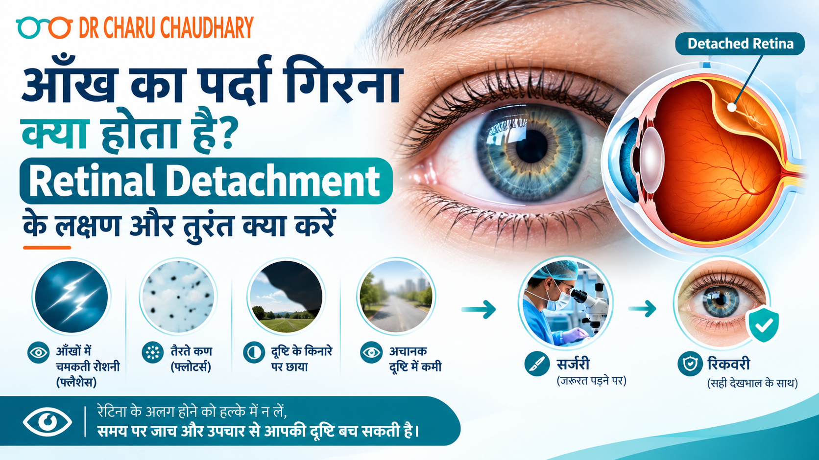
कल्पना कीजिए – आप सुबह उठते हैं और अचानक महसूस होता है कि एक आँख के सामने काला पर्दा गिर रहा है। या शायद रात में आपको बिजली जैसी चमक दिखी और सोचा “थकान होगी।” अगले दिन उसी आँख से धुंधला दिखने लगा। अधिकतर लोग इसे नज़रअंदाज़ कर देते हैं। यही सबसे बड़ी गलती है। जो आप महसूस कर रहे थे, वह Retinal Detachment – यानी आँख का पर्दा गिरना – के शुरुआती संकेत थे। और यह एक ऐसी medical emergency है जिसमें हर घंटा महत्वपूर्ण होता है। मैं Dr. Charu Chaudhary हूँ, Lucknow के Medanta Hospital में Vitreoretinal Surgeon। अपने वर्षों के अनुभव में मैंने देखा है कि जो मरीज़ 24–48 घंटे के भीतर आते हैं, उनकी रोशनी बचाई जा सकती है। जो देर करते हैं – उनके लिए रास्ता बहुत मुश्किल हो जाता है। इस blog में मैं आपको सरल भाषा में बताऊँगी कि आँख का पर्दा गिरना क्या होता है, इसके लक्षण क्या हैं, और सबसे ज़रूरी – इस स्थिति में आपको तुरंत क्या करना चाहिए। रेटिना क्या होती है और वह इतनी ज़रूरी क्यों है? आँख को एक camera की तरह समझिए। जिस तरह camera में film होती है जो तस्वीर कैद करती है – उसी तरह आँख में retina होती है। यह एक पतली, नाज़ुक परत होती है जो आँख के पिछले हिस्से में होती है। जब भी कोई रोशनी आँख में आती है, रेटिना उसे electrical signals में बदलता है और इन signals को Optic Nerve के ज़रिए दिमाग तक पहुँचाता है। दिमाग इन्हें image के रूप में पहचानता है – और आपको दिखता है। अगर यही रेटिना अपनी जगह से हट जाए – तो signal टूट जाता है। दृष्टि धुंधली हो जाती है, गायब हो सकती है, और बिना इलाज के हमेशा के लिए जा सकती है। आँख का पर्दा गिरता क्यों है – मुख्य कारण Retinal Detachment अचानक होता है, लेकिन इसके पीछे कुछ जोखिम कारण होते हैं जिनके बारे में आपको पहले से जानकारी होनी चाहिए: चोट या Trauma: आँख पर तेज़ चोट लगने से, पटाखे फूटने से, या खेल के दौरान चोट लगने से रेटिना खिंच सकती है और अपनी जगह से हट सकती है। High Myopia (तेज़ चश्मा): जिन लोगों का नंबर -6 या उससे अधिक है, उनकी आँख का आकार सामान्य से बड़ा होता है। इससे रेटिना पर खिंचाव रहता है और detachment का खतरा बढ़ जाता है। Diabetes: लम्बे समय से बेकाबू शुगर रेटिना की नसों को कमज़ोर करती है। इसे Diabetic Retinopathy कहते हैं – और यह Retinal Detachment का एक बड़ा कारण बन सकती है। बढ़ती उम्र: उम्र के साथ आँख के अंदर का gel (Vitreous) सिकुड़ता है और रेटिना को खींच सकता है। 50 वर्ष से अधिक उम्र के लोगों में यह ज़्यादा देखा जाता है। पहले का Retinal Detachment: एक आँख में हो चुका है तो दूसरी आँख में भी जोखिम बढ़ जाता है। पारिवारिक इतिहास: परिवार में किसी को हो चुका है तो आपको नियमित जाँच करानी चाहिए। आँख का पर्दा गिरने के लक्षण – जिन्हें कभी नज़रअंदाज़ न करें यह वह हिस्सा है जिसे मैं सबसे ज़्यादा ज़ोर देकर बताना चाहती हूँ। Retinal Detachment में दर्द नहीं होता—यही इसे इतना खतरनाक बनाता है। लोग सोचते हैं “दर्द नहीं तो गंभीर नहीं होगा।” यह सोच गलत है। इन लक्षणों में से कोई भी दिखे – तुरंत doctor के पास जाएँ: अचानक बहुत सारे काले धब्बे या जाले दिखना (Floaters) हाँ, कुछ floaters सामान्य हो सकते हैं। लेकिन अगर अचानक बहुत सारे नए floaters एक साथ दिखें – जैसे मकड़ी का जाला या काली बिंदियाँ तैर रही हों – तो यह रेटिना में खिंचाव या टूटन का संकेत है। बिजली जैसी चमक दिखना (Flashes of Light) आँखें बंद करने पर या अंधेरे में भी चमक दिखे – जैसे camera flash हो। यह इस बात का संकेत है कि Vitreous रेटिना को खींच रहा है। आँख के सामने काला पर्दा या छाया आना ऐसा महसूस हो जैसे किसी ने एक तरफ से curtain खींच दिया हो। पहले side vision जाती है, फिर धीरे-धीरे बीच का हिस्सा भी ढकने लगता है। यह सबसे serious लक्षण है। इस स्थिति में हर मिनट मायने रखता है। अचानक धुंधला दिखना, सिर्फ एक आँख से अचानक एक आँख से कम या धुंधला दिखना – और यह चश्मा लगाने पर भी ठीक न हो। यह इतनी बड़ी Emergency क्यों है? रेटिना एक जीवित tissue है। जब यह अपनी जगह से हट जाती है, तो उसे blood supply मिलनी बंद हो जाती है। जिस तरह खून न मिलने पर कोई भी अंग मरने लगता है – उसी तरह रेटिना की कोशिकाएँ भी मरने लगती हैं। पहले 24 घंटे: अगर पर्दा centre (Macula) तक नहीं पहुँचा, तो surgery के बाद अच्छी रोशनी वापस आने की संभावना बहुत अधिक है। 24–72 घंटे: Macula तक पहुँचने के बाद रोशनी वापस आना मुश्किल होने लगता है। 72 घंटे से अधिक: रेटिना की कोशिकाएँ स्थायी रूप से मर सकती हैं। रोशनी का नुकसान permanent हो सकता है। इसीलिए Retinal Detachment को हम “Golden Hour Emergency” कहते हैं। तुरंत क्या करें – Step by Step अगर आपको या आपके किसी परिवार वाले को ऊपर दिए गए लक्षण दिखें: Step 1: आँख को मलें नहीं, दबाएँ नहीं। Step 2: दोनों आँखें बंद करके शांत रहें – ज़्यादा eye movement से रेटिना खिंच सकता है। Step 3: कोई भी घरेलू उपाय, eye drop, या “देखते हैं कल” – यह सब बंद करें। Step 4: तुरंत किसी Vitreoretinal Specialist से मिलें। General physician या chemist के पास जाने का वक्त नहीं है। Lucknow में हैं तो अभी appointment book करें – हम emergency cases को उसी दिन देखते हैं। Diagnosis कैसे होती है? जब आप clinic आते हैं, तो हम कुछ ज़रूरी जाँचें करते हैं: Dilated Fundus Examination: आँख की पुतली को drops से बड़ा करके रेटिना को सीधे देखा जाता है। B-Scan Ultrasound: अगर आँख के अंदर खून है या साफ नहीं दिख रहा, तो Ultrasound से रेटिना की स्थिति देखी जाती है। OCT (Optical Coherence Tomography): रेटिना की परतों की detailed imaging – यह बताता है कि कितना नुकसान हुआ है। ये सब जाँचें दर्द रहित हैं
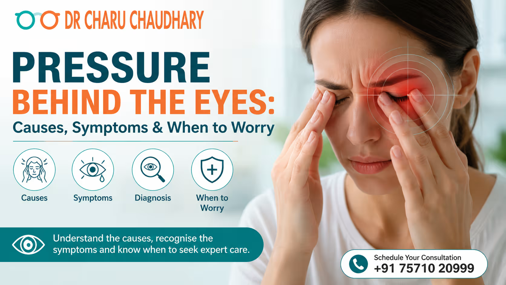
Pressure behind the eyes can feel like heaviness, aching, tightness, or a sensation that something is pushing from inside or around the eyes. Sometimes it is temporary and related to eye strain, sinus congestion, headaches, or fatigue. However, persistent or severe pressure—especially when accompanied by eye pain, redness, blurred vision, halos, nausea, or sudden vision changes—can indicate a condition that needs prompt medical evaluation. Importantly, the feeling of pressure behind the eyes does not always mean that your intraocular pressure (IOP) is high. High eye pressure often causes no noticeable symptoms and can only be identified through an eye examination and pressure measurement. If you frequently experience pressure behind your eyes, understanding the possible causes can help you know when it is time to consult an eye specialist. What Does Pressure Behind the Eyes Feel Like? People may describe eye pressure in different ways, including: The exact symptoms often depend on the underlying cause. Common Causes of Pressure Behind the Eyes 1. Eye Strain and Digital Eye Fatigue Long hours on computers, smartphones, or tablets can cause eye fatigue. Concentrating on a screen may also reduce blinking, contributing to dryness and discomfort. Common symptoms include: Taking regular screen breaks and adjusting screen distance, brightness, and lighting may help. 2. Sinus Congestion or Sinusitis The sinuses are located around the eyes, nose and forehead. Inflammation or congestion can create pressure or aching around these areas. Sinus pressure commonly occurs with colds, allergies, or sinus infections. You may also experience: If severe sinus pressure persists or is associated with significant symptoms, medical evaluation may be appropriate. 3. Migraine or Other Headaches Migraines can cause pain around or behind one eye. Some people also experience: The discomfort may occur even when there is no problem with the eye itself. 4. Dry Eyes Insufficient or poor-quality tears can cause irritation and a sensation of heaviness or pressure around the eyes. Typical symptoms include: Screen use, air conditioning, smoke and environmental factors can aggravate dry-eye symptoms. 5. Ocular Hypertension Ocular hypertension means that the pressure inside the eye is higher than normal without detectable optic nerve damage at that point. A key point is that ocular hypertension generally does not cause noticeable symptoms. It is usually discovered during an eye examination. This is why simply feeling pressure behind your eyes cannot tell you whether your eye pressure is actually elevated. 6. Glaucoma Glaucoma involves damage to the optic nerve. Increased eye pressure is an important risk factor, although glaucoma can also occur when eye pressure is within the usual range. Early glaucoma may have few or no symptoms. As the disease progresses, symptoms can include: Certain forms, particularly acute angle-closure glaucoma, can cause sudden severe symptoms and require immediate treatment. 7. Eye Inflammation or Infection Conditions affecting structures inside or around the eye can cause pain, pressure, redness and light sensitivity. Possible accompanying symptoms include: These symptoms should not be ignored, particularly when vision is affected. 8. Problems Around the Eye Socket Less commonly, inflammation, thyroid-related eye disease, injury, or other conditions affecting tissues behind the eye can produce pressure, discomfort or a feeling of fullness. Bulging of the eye, double vision, reduced vision, swelling, or difficulty moving the eye warrants medical assessment. Pressure Behind the Eyes: Cause vs Typical Symptoms Possible Cause Common Associated Symptoms Urgency Eye strain Tired eyes, headache, dryness Usually low Sinus pressure Blocked nose, facial pressure, headache Usually low–moderate Migraine Headache, nausea, light sensitivity Varies Dry eye Burning, gritty feeling, fluctuating vision Usually low Ocular hypertension Often no symptoms Needs eye evaluation Glaucoma Vision changes, peripheral vision loss Needs evaluation Acute angle-closure glaucoma Severe pain, red eye, halos, nausea Emergency Serious eye inflammation/injury Pain, redness, light sensitivity, blurred vision Prompt evaluation This comparison is educational; symptoms can overlap between conditions, so a proper eye examination is needed for diagnosis. When Is Pressure Behind the Eyes a Warning Sign? Seek urgent medical attention if pressure behind the eye occurs with: Acute angle-closure glaucoma can cause sudden eye pain or pressure, redness, blurred vision, halos and nausea/vomiting and is considered a medical emergency. Sudden vision loss, severe eye pain, or new flashes and floaters should also be assessed urgently. How Is Pressure Behind the Eyes Diagnosed? Because several conditions can produce similar sensations, an ophthalmologist may use multiple tests. Step 1: Medical and Eye History Your doctor may ask: Step 2: Comprehensive Eye Examination The doctor may examine the cornea, conjunctiva, pupils, lens, retina and optic nerve. Step 3: Measure Eye Pressure Tonometry measures intraocular pressure. Common methods include non-contact air-puff testing and contact-based measurements. Step 4: Check the Optic Nerve and Retina If glaucoma or another retinal/optic nerve condition is suspected, additional examinations may be recommended. Step 5: Additional Tests When Required Depending on your symptoms, your ophthalmologist may recommend: What Can You Do If You Have Mild Eye Pressure? If your symptoms are mild and there are no warning signs, you can consider these basic measures: Do not assume that recurring pressure is simply due to screen use, especially if you have vision changes. Can High Eye Pressure Cause a Feeling of Pressure? Not necessarily. This is one of the most important things to understand. High intraocular pressure often has no obvious symptoms. A person can have elevated IOP without feeling pain or pressure. The only reliable way to determine whether your eye pressure is elevated is through an appropriate eye examination and pressure measurement. Therefore: Feeling pressure ≠ automatically having high eye pressure. And: Having high eye pressure ≠ necessarily feeling pressure. This distinction is particularly important when screening for glaucoma. When Should You See an Eye Specialist? Consider scheduling an eye examination if: Regular eye examinations can help detect problems that may not cause symptoms in their early stages. Pressure Behind the Eyes: Quick Decision Guide Mild pressure + screen use + tired eyes→ Take breaks, rest your eyes and monitor symptoms. Pressure + blocked nose/facial congestion→ A sinus or allergy-related cause may be possible. Recurring pressure
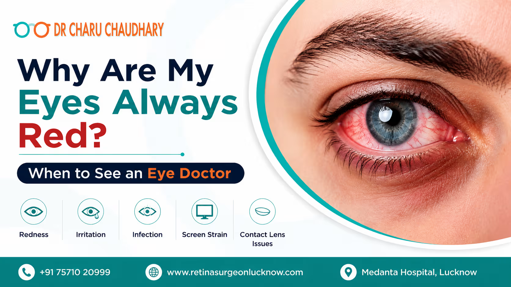
If your eyes are always red, the cause may be dry eyes, allergies, prolonged screen time, contact lens irritation, conjunctivitis, or another eye condition. Mild redness may improve with rest, but persistent redness—especially with pain, light sensitivity, discharge, or vision changes—should be evaluated by an eye doctor. Red eyes are one of the most common eye complaints. Sometimes they are temporary and harmless, such as after a sleepless night or long hours in front of a screen. However, eyes that remain red or become red repeatedly may indicate an underlying problem that needs proper treatment. Understanding the possible red eyes causes can help you know when simple eye care is enough and when you should consult an ophthalmologist. What Does Eye Redness Mean? Eye redness occurs when the small blood vessels on the surface of the eye become enlarged, irritated, or inflamed. This makes the normally white part of the eye appear pink or red. Eye redness can affect one or both eyes and may occur with other symptoms, including: The severity and combination of symptoms can provide clues about the possible cause. However, symptoms alone cannot always identify the problem. If your eyes are always red or frequently become red without an obvious reason, an eye examination can help determine the underlying cause. Common Red Eyes Causes There are many possible causes of eye redness. Some are minor and temporary, while others require medical treatment. 1. Dry Eyes Dry eye is a common reason for persistent redness. It can occur when the eyes do not produce enough tears or when tears evaporate too quickly. Dry eyes may cause: Long screen hours, reduced blinking, air conditioning, pollution and certain environmental conditions can contribute to dry eye symptoms. 2. Eye Allergies Allergic conjunctivitis can cause red, itchy and watery eyes. Common triggers include dust, pollen, pet dander, smoke and other allergens. Intense itching is particularly common with eye allergies. Allergy-related redness often affects both eyes. 3. Conjunctivitis or Pink Eye Conjunctivitis occurs when the conjunctiva becomes inflamed. Viruses, bacteria or allergies can cause it. Possible symptoms include: Viral and bacterial conjunctivitis can be contagious, so good hand hygiene and avoiding shared towels or cosmetics are important. 4. Prolonged Screen Time Long hours on computers, smartphones and other digital devices can contribute to digital eye strain. When concentrating on a screen, people often blink less frequently. Reduced blinking can destabilize the tear film and cause dryness and irritation, which may make the eyes look red. 5. Lack of Sleep Insufficient sleep can leave the eyes looking tired, dry and bloodshot. Occasional redness after a late night usually improves with adequate sleep and hydration. However, frequent redness should not automatically be blamed on lack of sleep. 6. Contact Lens Problems Contact lenses can cause redness when they are worn incorrectly or for longer than recommended. Poor lens hygiene, sleeping in unsuitable lenses, using expired lenses, improper cleaning or continuing to wear lenses despite irritation can increase the risk of eye-surface problems and infection. 7. Foreign Body or Eye Injury Dust, sand, chemicals or another foreign object can irritate the eye and cause sudden redness. Do not rub the eye because rubbing can worsen irritation or potentially damage the cornea. Chemical exposure to the eye requires prompt medical attention. 8. Other Eye Conditions In some cases, red eyes may be associated with more significant conditions involving the cornea, uvea, intraocular pressure or other eye structures. This is particularly important when redness occurs with pain, blurred vision, light sensitivity, nausea, halos around lights or other sudden changes in vision. Symptoms That May Point to the Cause The symptoms occurring alongside redness can provide useful clues, although only an eye examination can establish the diagnosis. Symptoms Possible Cause Redness + intense itching Eye allergy Redness + watery discharge Allergy or viral conjunctivitis Redness + thick discharge Possible bacterial conjunctivitis Redness + burning or grittiness Dry eye or irritation Redness + contact lens use Lens-related irritation or infection Redness + significant pain Requires prompt evaluation Redness + blurred vision Needs medical assessment Redness + light sensitivity May indicate a more significant eye problem Redness after an injury Possible eye trauma Redness + sudden vision changes Urgent eye assessment may be required These associations are only general indicators. Avoid self-diagnosing based on symptoms alone. Can Contact Lenses Cause Redness? Yes, contact lenses can cause eye redness, particularly when they are not used or maintained correctly. Common reasons include: Contact lens wearers should pay particular attention to painful red eyes. If you develop significant redness, pain, light sensitivity, discharge or reduced vision while wearing contact lenses, remove the lenses and seek professional eye care promptly. Do not simply use redness-relieving drops and continue wearing the lenses. Eye Allergy or Eye Infection: How Are They Different? Eye allergies and eye infections can produce similar symptoms, making it difficult to distinguish between them without an examination. Eye Allergy Eye allergies commonly cause: Symptoms may worsen after exposure to dust, pollen, smoke, pets or other allergens. Eye Infection An eye infection may cause: Viral conjunctivitis often causes watery discharge, while bacterial conjunctivitis may produce thicker discharge. However, these symptoms can overlap. Antibiotic eye drops should not be started simply because the eye is red, because antibiotics do not treat viral or allergic causes. An ophthalmologist can determine the likely cause and recommend appropriate treatment. Safe Care for Mild Redness For mild redness without severe pain, vision changes or other warning signs, simple measures may help reduce irritation. Helpful steps include: Avoid these common mistakes Do not repeatedly use eye drops marketed only to “remove redness” without understanding the cause. Temporary whitening of the eye does not necessarily mean the underlying problem has been treated. Also avoid using leftover antibiotic or steroid eye drops without an ophthalmologist’s advice. Some eye conditions can worsen when inappropriate medication is used. When to See an Eye Doctor Not every red eye is an emergency, but persistent or recurrent eye redness should be evaluated if it
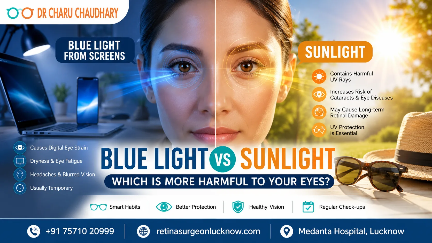
Have you ever wondered whether spending hours on your phone is more harmful than walking under the bright sun? This is one of the most common concerns people discuss with an eye specialist in Lucknow, especially as screen time continues to increase in both work and daily life. While smartphones, laptops, and tablets emit blue light, sunlight is the largest natural source of blue light and also contains harmful ultraviolet (UV) rays. Understanding the difference is essential for protecting your eyesight. The truth is that normal screen use is more likely to cause temporary eye strain and dryness, whereas prolonged exposure to sunlight without proper protection poses a greater long-term risk to eye health because of UV radiation. Knowing how each affects your eyes can help you adopt healthier habits and reduce your risk of vision problems. In this comprehensive guide, we’ll explain the science behind blue light and sunlight, separate myths from facts, and share expert-recommended strategies to keep your eyes healthy. Understanding Blue Light vs Sunlight Blue light is a part of the visible light spectrum, with wavelengths ranging from approximately 400 to 500 nanometers. It is often referred to as high-energy visible (HEV) light because it has a shorter wavelength and higher energy than most other visible colors. Many people associate blue light only with smartphones or computers, but in reality, the sun is by far the strongest natural source of blue light. Digital devices emit only a small fraction of the blue light that reaches your eyes outdoors. Common Sources of Blue Light Natural sources include: Artificial sources include: Although both sunlight and digital devices emit blue light, their intensity, duration, and accompanying radiation differ significantly, which is why they affect the eyes in different ways. Why Blue Light Matters Not all blue light is harmful. In fact, natural blue light plays an important role in maintaining overall health. Benefits of Natural Blue Light Natural blue light helps: Without exposure to natural daylight, our biological clock can become disrupted, affecting sleep quality and overall well-being. However, excessive exposure to intense light—particularly sunlight without adequate eye protection – may increase the risk of certain eye conditions over time. How the Eye Processes Light Before understanding which type of light is more harmful, it’s helpful to know how light travels through the eye. When light enters the eye, it passes through: The retina converts light into electrical signals, which are transmitted through the optic nerve to the brain, allowing us to see. The eye has natural protective mechanisms that filter part of the blue light reaching the retina. However, ultraviolet (UV) rays from sunlight interact differently with eye tissues and can contribute to long-term damage when exposure is excessive. Why Your Eyes Respond Differently to Blue Light and Sunlight Many people assume that all blue light is equally harmful, but this is a misconception. The way your eyes respond depends on the intensity of the light source, duration of exposure, and whether ultraviolet (UV) radiation is present. Blue Light From Digital Screens Blue light emitted by digital screens is: This is why people often experience symptoms such as dry eyes, headaches, and blurred vision after long hours of screen use. Sunlight Sunlight contains: Unlike screens, sunlight exposes your eyes to significantly higher energy levels and harmful UV radiation. Over many years, unprotected UV exposure may increase the risk of: This is why ophthalmologists recommend wearing UV400 sunglasses whenever you spend extended time outdoors. Expert Insight One of the biggest misconceptions is that mobile phones permanently damage the retina simply because they emit blue light. Current ophthalmic research indicates that the amount of blue light produced by smartphones and computers is substantially lower than that from natural sunlight. For most people, the symptoms experienced after prolonged screen use are related to digital eye strain, reduced blinking, glare, and poor ergonomics, rather than retinal injury. Blue Light vs Sunlight: Key Differences The following comparison highlights why sunlight and digital screens affect your eyes differently. Feature Blue Light From Screens Sunlight Primary Source Smartphones, tablets, laptops, LED screens Natural sunlight Blue Light Intensity Low Very High Contains UV Rays No Yes Causes Digital Eye Strain Yes Rarely Can Cause Dry Eyes Yes No Long-Term UV Damage No Yes Risk of Cataracts No direct link Increased with prolonged UV exposure Risk to Retina No proven damage from normal use Direct sun viewing can damage the retina Best Protection Screen breaks, blinking, ergonomic setup UV400 sunglasses, hats, avoiding direct sun Does Blue Light From Screens Harm the Retina? One of the most frequently searched questions online is whether blue light from smartphones permanently damages the retina. The current scientific evidence provides reassuring answers. Studies have not demonstrated that normal exposure to blue light from digital devices causes permanent retinal damage in healthy individuals. The amount of blue light emitted by screens is significantly lower than that produced by natural sunlight. Most people who spend long hours using digital devices experience symptoms related to Digital Eye Strain (Computer Vision Syndrome) rather than retinal disease. Common Symptoms Associated with Prolonged Screen Use These symptoms usually improve with proper screen habits, regular breaks, and appropriate lighting. However, individuals with pre-existing retinal conditions, such as diabetic retinopathy or age-related macular degeneration, should follow the advice of their ophthalmologist regarding prolonged screen use. What Causes Digital Eye Strain? One of the biggest misconceptions is that blue light itself is the main reason for eye discomfort after using digital devices. In reality, ophthalmologists agree that Digital Eye Strain, also known as Computer Vision Syndrome (CVS), is usually caused by the way we use screens rather than the blue light they emit. Whether you’re working on a computer, attending online classes, or scrolling through social media, your eyes work harder to maintain focus on digital screens than they do when reading printed text. This constant effort, combined with poor viewing habits, can lead to a range of uncomfortable symptoms. Why Does Digital Eye Strain Happen? Several factors contribute to
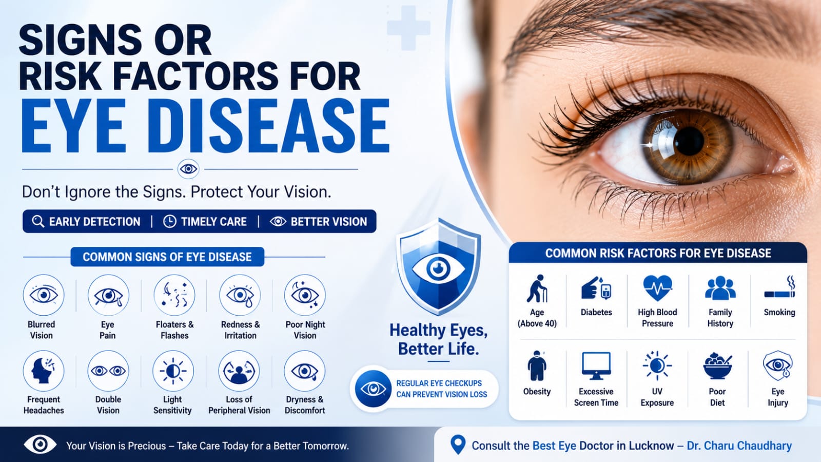
Signs and Risk Factors for Eye Disease Our eyes help us experience the world every day, yet many people ignore early symptoms of eye disease until the condition becomes serious. The truth is that many eye diseases develop slowly and may not cause pain in the beginning. Detecting them early can help protect your vision and prevent permanent damage. Whether you are young or old, understanding the warning signs and risk factors can help you take better care of your eyesight. If you notice any unusual changes in your vision, consulting Dr. Charu Chaudhary, widely regarded as the best eye specialist in Lucknow, can help identify problems before they become severe. Why Early Detection of Eye Disease Matters Many eye diseases do not show noticeable symptoms in their early stages. Conditions like glaucoma, diabetic retinopathy, and age-related macular degeneration can silently damage vision over time. Early diagnosis allows: Regular eye examinations remain the best way to detect these diseases before symptoms become severe. Common Signs of Eye Disease Recognizing the warning signs can save your eyesight. 1. Blurred Vision Blurred vision that does not improve with blinking may indicate: 2. Sudden Vision Loss Any sudden decrease in vision is a medical emergency and requires immediate attention. Possible causes include: 3. Eye Pain Pain inside or around the eye should never be ignored. It may indicate: 4. Frequent Headaches Persistent headaches accompanied by eye strain may result from: 5. Double Vision Seeing two images instead of one may indicate neurological or eye muscle disorders. 6. Floaters and Flashes Occasional floaters are common with age, but a sudden increase in floaters or flashes of light can signal retinal tears. 7. Red Eyes Redness lasting more than a day could indicate: 8. Difficulty Seeing at Night Poor night vision may be caused by: 9. Light Sensitivity Excessive sensitivity to light may indicate: 10. Loss of Peripheral Vision This is often one of the earliest signs of glaucoma. Common Risk Factors for Eye Disease Several factors increase your chances of developing eye disorders. Risk Factor Why It Increases Risk Age above 40 Higher risk of cataracts and glaucoma Diabetes Can damage retinal blood vessels High Blood Pressure Affects blood supply to the eyes Family History Many eye diseases are hereditary Smoking Increases cataract and macular degeneration risk Obesity Raises risk of diabetic eye disease Excessive Screen Time Causes eye strain and dry eyes UV Exposure Damages the lens and retina Poor Diet Lack of vitamins affects eye health Eye Injury Can lead to long-term complications Eye Diseases and Their Early Symptoms Eye Disease Early Symptoms Cataract Blurred vision, glare, poor night vision Glaucoma Peripheral vision loss, eye pressure Diabetic Retinopathy Blurred vision, floaters Dry Eye Syndrome Burning, irritation, redness Macular Degeneration Distorted central vision Retinal Detachment Flashes, floaters, sudden vision loss Conjunctivitis Redness, discharge, itching Who Should Get Regular Eye Checkups? You should schedule regular eye examinations if you: Routine checkups help detect hidden eye diseases before symptoms appear. How to Reduce Your Risk of Eye Disease Eat an Eye-Healthy Diet Include foods rich in: Good choices include carrots, spinach, kale, fish, eggs, citrus fruits, almonds, and sweet potatoes. Protect Your Eyes from UV Rays Wear sunglasses that block 100% UVA and UVB rays whenever outdoors. Control Diabetes and Blood Pressure Keeping blood sugar and blood pressure under control significantly reduces the risk of diabetic eye disease. Follow the 20-20-20 Rule If you use digital devices frequently: Every 20 minutes, Look 20 feet away, For at least 20 seconds. This helps reduce digital eye strain. Quit Smoking Smoking damages blood vessels in the eyes and increases the risk of cataracts and macular degeneration. Exercise Regularly Regular physical activity improves blood circulation and lowers the risk of chronic diseases that affect vision. When Should You Visit an Eye Doctor? Seek immediate medical attention if you experience: Ignoring these symptoms may result in permanent vision damage. Why Choose Dr. Charu Chaudhary for Eye Care? Choosing the right eye specialist plays an important role in protecting your vision. Dr. Charu Chaudhary is known for providing comprehensive eye care with advanced diagnostic techniques and personalized treatment plans. Whether you need a routine eye examination, cataract consultation, glaucoma screening, diabetic eye evaluation, or treatment for other eye conditions, patients trust her expertise and compassionate care. If you are searching for the best eye doctor in Lucknow, Dr. Charu Chaudhary offers modern eye care focused on early diagnosis, accurate treatment, and long-term vision protection. Frequently Asked Questions (FAQs) What are the first signs of eye disease? Blurred vision, eye pain, flashes of light, floaters, redness, and loss of peripheral vision are among the earliest warning signs. Can eye diseases be prevented? Many eye diseases cannot be completely prevented, but regular eye exams, a healthy lifestyle, and early treatment can significantly reduce the risk of vision loss. How often should adults have an eye examination? Adults should have a comprehensive eye examination every one to two years. People with diabetes, glaucoma risk, or existing eye problems may require more frequent checkups. Does screen time permanently damage eyesight? Screen time generally does not cause permanent vision damage but can lead to digital eye strain, dry eyes, headaches, and temporary blurred vision. Why should diabetic patients have regular eye checkups? Diabetes can silently damage the retina without early symptoms. Regular eye exams help detect diabetic retinopathy before permanent vision loss occurs. Conclusion Your eyesight is one of your most valuable assets. Many eye diseases begin silently, making regular eye examinations essential for early diagnosis and effective treatment. Recognizing warning signs such as blurred vision, eye pain, floaters, flashes of light, or persistent redness can help prevent serious complications. If you notice any of these symptoms or have risk factors like diabetes, high blood pressure, or a family history of eye disease, don’t delay seeking professional care. Dr. Charu Chaudhary, recognized by many patients as the best eye doctor in Lucknow, provides comprehensive eye care to help protect your vision and maintain lifelong
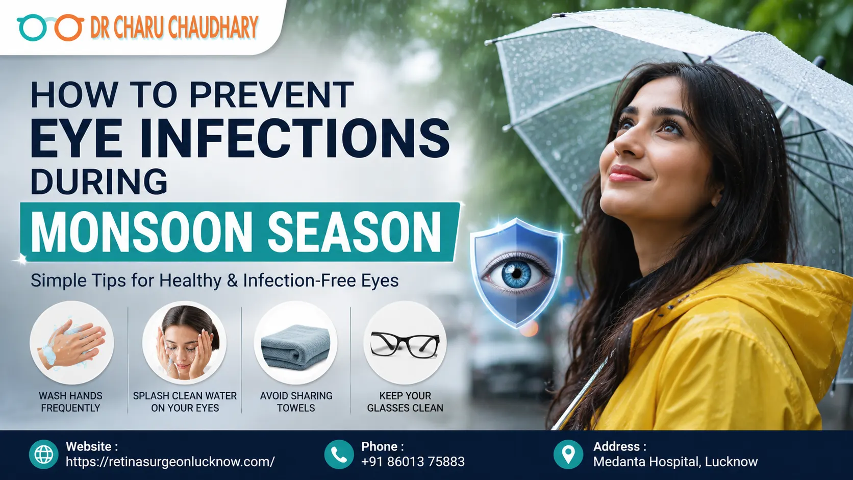
The monsoon season brings welcome relief from the summer heat, but it also creates ideal conditions for bacteria, viruses, fungi, and allergens to thrive. Increased humidity, stagnant water, and poor hygiene significantly raise the risk of eye infections. From viral conjunctivitis to styes and allergic eye irritation, monsoon-related eye problems can affect people of all ages. Fortunately, most eye infections are preventable with proper hygiene and timely care. In this comprehensive guide, you’ll learn how to prevent eye infections during monsoon season, recognize early warning signs, understand available treatments, and know when to consult the Eye Surgeon in Lucknow. Why Do Eye Infections Increase During the Monsoon? The rainy season creates an environment that promotes the growth and spread of microorganisms. Some common reasons include: Children, elderly individuals, contact lens users, diabetic patients, and people with weakened immunity are particularly vulnerable. Common Eye Infections During Monsoon 1. Viral Conjunctivitis (Pink Eye) One of the most common monsoon eye infections. Symptoms It spreads easily through direct contact and contaminated objects. 2. Bacterial Conjunctivitis Unlike viral conjunctivitis, bacterial infections often produce thick yellow or green discharge. Symptoms include: Prompt treatment prevents complications. 3. Stye (Eye Boil) A stye is a painful infection of an eyelid gland. Symptoms include: Poor eyelid hygiene increases the risk. 4. Fungal Eye Infection Although less common, fungal infections may occur after eye injuries involving contaminated water or soil. Symptoms: These infections require urgent ophthalmic evaluation. 5. Allergic Eye Disease Monsoon humidity increases mold spores and allergens. Symptoms: Unlike infections, allergies are not contagious. Signs You May Have an Eye Infection Watch for these warning symptoms: Never ignore sudden vision loss or severe eye pain. How to Prevent Eye Infections During Monsoon Season Preventive care is always better than treatment. Follow these practical eye care tips throughout the rainy season. 1. Wash Your Hands Frequently Hands carry thousands of germs every day. Always wash your hands: Use soap and clean water for at least 20 seconds. 2. Avoid Touching or Rubbing Your Eyes Eye rubbing transfers bacteria and viruses directly to the eye surface. Instead: 3. Keep Your Eyes Clean Clean your eyelids gently using: Avoid using dirty towels. 4. Never Share Personal Items Avoid sharing: Many eye infections spread through shared items. 5. Maintain Contact Lens Hygiene Contact lens users have a higher infection risk. Always: Stop wearing lenses immediately if your eyes become red or painful. 6. Protect Your Eyes from Rainwater Rainwater is not always clean. If rainwater enters your eyes: 7. Wear Protective Glasses Outdoors Protective eyewear helps prevent: Sunglasses also reduce irritation from wind. 8. Eat Eye-Healthy Foods Strong immunity reduces infection risk. Include: Vitamin A, Vitamin C, Vitamin E, zinc, and antioxidants support healthy eyes. 9. Stay Hydrated Even during rainy weather, dehydration can affect tear production. Drink: Good hydration supports healthy tear film function. 10. Avoid Self-Medication Never use: Improper treatment may worsen the infection. Eye Care Tips for Children During Monsoon Children frequently catch viral conjunctivitis. Parents should: Early treatment prevents spread within families and schools. What to Do if You Develop an Eye Infection If symptoms appear: Do Don’t When Should You Visit an Eye Specialist? Seek immediate medical attention if you experience: Prompt diagnosis helps prevent serious complications. Can Eye Infections Affect Vision? Most mild infections resolve completely with proper treatment. However, untreated infections can lead to: Early diagnosis greatly reduces these risks. How Eye Specialists Diagnose Eye Infections During your eye examination, your ophthalmologist may perform: Accurate diagnosis ensures appropriate treatment. Simple Daily Monsoon Eye Care Routine Follow this checklist every day: Consistency is the key to preventing seasonal eye infections. Conclusion Monsoon season increases the risk of eye infections, but simple preventive measures can keep your eyes healthy. Maintaining good hygiene, avoiding contaminated water, practicing safe contact lens care, and recognizing early symptoms can significantly reduce your chances of developing infections. If redness, pain, discharge, or blurred vision develops, don’t ignore the symptoms. Early diagnosis and timely treatment protect your eyesight and prevent complications. Consulting the Best Eye Specialist in Lucknow ensures you receive an accurate diagnosis and advanced treatment tailored to your condition.
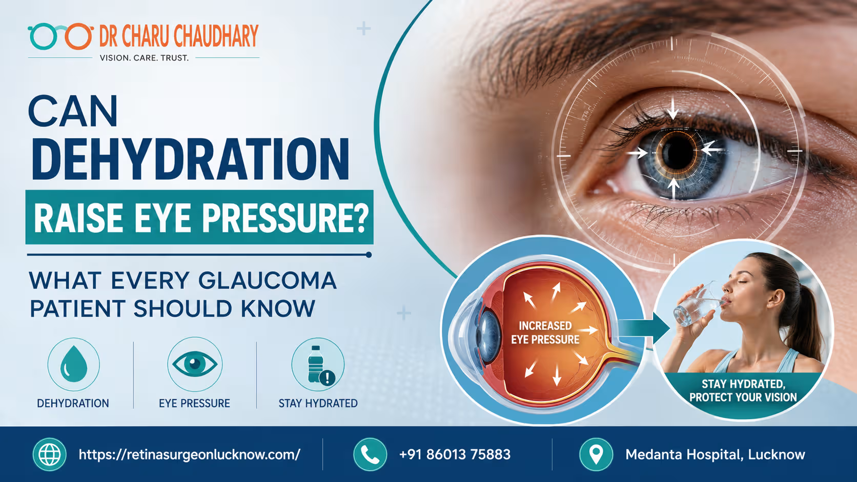
Dehydration may temporarily influence intraocular pressure in some people and can significantly affect overall eye health. While dehydration alone does not usually cause glaucoma, maintaining proper hydration is important for patients already diagnosed with the condition. Fluctuations in body fluids can impact how the eye regulates its internal pressure and supports the optic nerve. Can Not Drinking Enough Water Affect Your Eye Pressure? Maintaining the right fluid balance is vital for your health. Most people know that dehydration affects the heart and kidneys. However, many patients are surprised to learn that it also affects their eyes. For individuals living with glaucoma, understanding the link between hydration and eye pressure is essential. Eye pressure, or intraocular pressure (IOP), is the primary metric used to manage glaucoma. When your body lacks sufficient water, your systemic circulation changes. These changes can impact the delicate structures inside your eye. Consequently, your vision may face unnecessary risks if you ignore your daily water intake. Dr. Charu Chaudhary, an expert in ocular health, emphasizes that hydration is a pillar of preventative care. She often advises that stable fluid levels help maintain a stable environment for the optic nerve. If you are looking for the Best Eye Specialist in Lucknow, finding a professional who looks at your systemic health is key. Why Glaucoma Patients Should Pay Attention to Hydration Glaucoma is often called the “silent thief of sight.” It slowly damages the optic nerve, usually due to high internal eye pressure. Because this condition is so sensitive to pressure changes, even minor fluctuations matter. Proper hydration ensures that the blood flowing to your eyes is rich in oxygen and nutrients. When you are dehydrated, your blood can become more viscous. This change may reduce the efficiency of blood flow to the optic nerve. Therefore, staying hydrated isn’t just about the “pressure number” on a chart. It is about protecting the nerve fibers that allow you to see. Is there a real connection between dehydration and eye health? Medical research suggests that while severe dehydration might actually lower eye pressure slightly, the secondary effects are harmful. Dry eyes, blurred vision, and poor circulation can worsen the overall experience of living with glaucoma. What Is Eye Pressure (Intraocular Pressure)? Intraocular pressure is the fluid pressure inside your eye. Your eye produces a clear fluid called aqueous humor. This fluid nourishes the front part of the eye and maintains its shape. To keep pressure steady, the eye must drain as much fluid as it produces. When the drainage system becomes clogged or inefficient, the fluid builds up. This buildup increases the pressure within the eyeball. Think of it like a balloon being overfilled with water. If the pressure remains too high for too long, it pushes against the back of the eye. How Is Eye Pressure Measured? Eye specialists use a process called tonometry to measure IOP. During an exam, the doctor will use a device to gently touch or blow a puff of air onto the surface of your eye. This measurement is recorded in millimeters of mercury (mmHg). A normal eye pressure range is generally between 12 and 21 mmHg. However, “normal” can vary from person to person. Some people develop glaucoma with low pressure, while others have high pressure without nerve damage. This is why regular monitoring is so crucial for long-term vision. Why Is Eye Pressure Important for Vision? Your optic nerve is the “cable” that connects your eye to your brain. It is made of over a million tiny nerve fibers. High eye pressure compresses these fibers. Over time, the fibers begin to die off, leading to permanent vision loss. Because these fibers do not regenerate, the damage is irreversible. Managing eye pressure is the only proven way to stop or slow the progression of glaucoma. Consistency in your habits, including your water intake, helps keep these pressure levels within a safe range. What Is Glaucoma and How Does It Damage Vision? Glaucoma is a group of eye conditions that lead to optic nerve damage. It is typically categorized by how the fluid drainage system is blocked. The most common form is Open-Angle Glaucoma, where the drainage angle looks open but functions poorly. Another form is Angle-Closure Glaucoma, which can occur suddenly and constitutes a medical emergency. Regardless of the type, the result is often the same: peripheral vision fades first. If left untreated, the damage moves inward, eventually leading to total blindness. High eye pressure is the most significant modifiable risk factor for this disease. By controlling this pressure through medication, surgery, and lifestyle choices, patients can preserve their sight for a lifetime. This is why consulting the Best Eye Surgeon in Lucknow is a vital step for those at risk. Can Dehydration Really Raise Eye Pressure? The relationship between dehydration and eye pressure is complex. In many cases, severe systemic dehydration actually leads to a decrease in intraocular pressure. This happens because the body produces less aqueous humor when it is trying to conserve water. However, the problem arises when the body is in a state of flux. Chronic dehydration can lead to “dry eye” symptoms that mimic or worsen glaucoma discomfort. Furthermore, when a dehydrated person suddenly drinks a massive amount of water, it can cause a temporary spike in eye pressure. What Does Medical Research Say? Research indicates that the body’s “osmolality”—the concentration of particles in your blood—affects the fluid in your eyes. When you are dehydrated, your blood is more concentrated. This can pull fluid out of the eye, lowering pressure. Conversely, “water loading” (drinking a liter of water in under five minutes) is a known test to see if eye pressure will spike. For a glaucoma patient, these rapid shifts are dangerous. Stability is the goal. Therefore, avoiding extreme dehydration is just as important as avoiding rapid over-hydration. Is the Effect Temporary or Long-Term? Most pressure changes related to hydration are temporary. However, the cumulative effect of poor hydration on the optic nerve’s blood supply can be long-term. Your eyes need consistent hydration
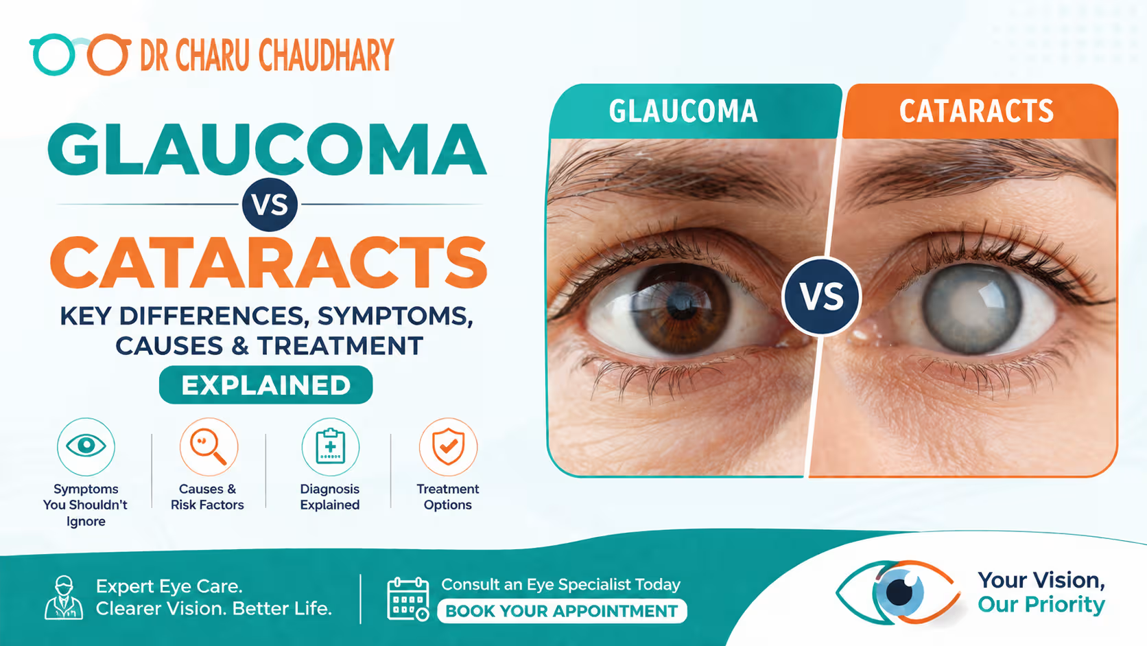
Cataracts cause cloudy vision because the eye’s natural lens becomes opaque, blocking light from reaching the retina. Conversely, glaucoma damages the optic nerve, often due to increased eye pressure. While cataract surgery usually restores vision successfully, glaucoma-related vision loss is irreversible. Therefore, early diagnosis is essential to prevent permanent blindness. Introduction How Can You Tell Whether It’s Glaucoma or Cataracts? Distinguishing between different eye conditions is often difficult for the average person. You might notice your vision becoming blurry or find it hard to drive at night. However, these symptoms could point toward two very different diseases. Glaucoma and cataracts are the leading causes of vision impairment globally. While they share some similarities, their impact on your eye health varies significantly. Identifying the specific cause of your visual decline is the first step toward effective recovery. Why Do People Often Confuse These Two Eye Conditions? Confusion often arises because both conditions commonly affect older adults. Furthermore, both diseases can cause a gradual decline in visual clarity. Patients frequently assume that any blurring of vision is simply a sign of “getting older.” Unfortunately, this misconception can lead to delayed treatment. While one condition is often fixable, the other can lead to permanent darkness if ignored. Understanding the biological differences between them is crucial for every patient. Why Early Eye Examination Can Protect Your Vision Regular screenings are the only definitive way to distinguish between these two conditions. An experienced Eye doctor in Lucknow can perform a comprehensive evaluation to assess your ocular health. Early detection allows for a wider range of treatment options. Most importantly, it helps prevent the silent progression of diseases that could steal your sight. By scheduling an exam, you take a proactive step in preserving your quality of life. What Is Glaucoma and How Does It Affect Vision? Glaucoma is not a single disease but a group of eye conditions. These conditions primarily damage the optic nerve, which is vital for good vision. This damage is often caused by abnormally high pressure inside your eye. Over time, this pressure erodes the nerve fibers, leading to blind spots in your visual field. What Happens to the Optic Nerve? The optic nerve acts like a communication cable between your eye and your brain. It carries thousands of electrical impulses that the brain interprets as images. When intraocular pressure rises, it compresses these delicate nerve fibers. Consequently, the fibers begin to die, and the “cable” loses its ability to transmit information. This process usually starts with your side vision and slowly moves inward. Why Is Glaucoma Called the “Silent Thief of Sight”? Many forms of glaucoma have no warning signs. For example, open-angle glaucoma progresses so slowly that you may not notice a change until the late stages. The brain often compensates for small blind spots, making the disease nearly invisible to the patient. By the time vision loss becomes obvious, significant and permanent damage has already occurred. This is why medical professionals emphasize the importance of preventative screenings. What Are Cataracts and Why Do They Develop? A cataract is a clouding of the normally clear lens of your eye. For people who have cataracts, seeing through cloudy lenses is a bit like looking through a frosty window. This clouding makes it more difficult to read or drive a car, especially at night. Most cataracts develop slowly and don’t disturb your eyesight early on. How Does the Eye Lens Become Cloudy? The lens is positioned behind the colored part of your eye (the iris). It focuses light that passes into your eye, producing clear, sharp images on the retina. As you age, the proteins within the lens begin to break down and clump together. These clumps create a cloudy area that scatters light instead of focusing it clearly. As a result, the image reaching your retina becomes blurred or distorted. Who Is Most Likely to Develop Cataracts? Aging is the most significant risk factor for cataract development. Most people begin to develop some degree of lens clouding after the age of 40. However, other factors can accelerate this process significantly. These include prolonged exposure to ultraviolet (UV) light, smoking, and certain medications like steroids. Additionally, individuals with systemic health issues may see symptoms appear much earlier in life. What Is the Difference Between Glaucoma and Cataracts? While both conditions affect your ability to see, they involve different parts of the eye. Cataracts are a problem with the “lens,” which is the front window of the eye. Glaucoma is a problem with the “nerve,” which is the connection to the brain. Understanding this fundamental difference helps patients appreciate why the treatments differ so vastly. How Do Their Causes Differ? The causes of these two diseases are entirely unrelated. Cataracts are primarily a result of protein degradation and oxidative stress within the lens. In contrast, glaucoma usually stems from a failure in the eye’s fluid drainage system. When the aqueous humor (eye fluid) cannot drain properly, pressure builds up against the retina and optic nerve. Therefore, one is a structural change in a tissue, while the other is often a hydraulic issue. How Do Symptoms Compare? Cataract symptoms usually involve a change in how you perceive light and color. You might see “halos” around streetlights or find that colors look faded. Glaucoma symptoms are often non-existent in the early stages. However, advanced glaucoma leads to “tunnel vision,” where your peripheral sight disappears. While cataracts make the world look “fuzzy,” glaucoma makes the world look “smaller.” Which Condition Progresses Faster? Progression rates vary greatly between individuals and the specific type of the disease. Cataracts generally progress very slowly over several years. Many patients live with mild cataracts for a long time without needing surgery. Some forms of glaucoma, such as acute angle-closure glaucoma, can cause vision loss within hours. Chronic glaucoma, however, also moves slowly but leaves permanent damage in its wake. Glaucoma vs. Cataract Comparison Table Feature Glaucoma Cataracts Primary Site Optic Nerve Crystalline Lens Main Cause High Intraocular Pressure Protein Clumping/Aging
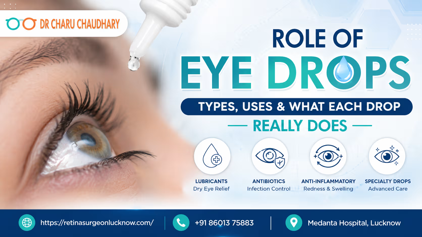
When your eyes feel itchy, dry, or red, the first thing most people reach for is a bottle of eye drops. However, walking into a pharmacy can be overwhelming. There are hundreds of bottles, each claiming to solve a different problem. Are they all the same? The short answer is no. Using the wrong eye drop can not only be ineffective but, in some cases, dangerous. What do eye drops actually do?Different eye drops are designed for specific medical purposes: lubricating drops soothe dry eyes, antibiotic drops fight bacterial infections, allergy drops block histamine, and glaucoma drops lower intraocular pressure. They also reduce inflammation, aid post-surgical recovery, or dilate pupils, ensuring targeted delivery of medication directly to ocular tissues. If you are looking for an eye doctor in Lucknow, understanding the nuances of ocular medication is the first step toward better vision health. This guide will walk you through everything you need to know about the role of eye drops. Introduction Why So Many People Use Eye Drops Without Understanding Them The eye is one of the most accessible organs for topical treatment. Because eye drops are available over the counter (OTC), many patients treat them like lip balm or moisturizer—something to use whenever there is slight discomfort. However, the eye is also incredibly delicate. Applying a liquid to the surface of the eye isn’t just about “washing” it; it’s about introducing active pharmacological agents into a complex biological system. Are All Eye Drops the Same? Far from it. While most eye drops look like clear water, their chemical compositions vary wildly. A drop designed to constrict blood vessels to “get the red out” works entirely differently than a drop designed to lower internal eye pressure for glaucoma. Confusing the two can lead to missed diagnoses and permanent vision loss. Why Choosing the Right Eye Drop Matters Using an antibiotic drop for a viral infection is useless. Using a steroid drop for a simple scratch on the cornea can lead to a fungal infection or a non-healing ulcer. As Dr Charu Chaudhary, a renowned Eye Specialist in Lucknow, often emphasizes, “The eye does not have a high tolerance for the wrong medication. Precision in diagnosis is the only way to ensure safety.” Understanding How Eye Drops Work How Medication Reaches the Eye When you swallow a pill, the medication must travel through your digestive system, into your bloodstream, and eventually find its way to your eyes. The “blood-ocular barrier” makes this difficult, meaning high doses of oral medicine are often needed to treat the eyes. Eye drops bypass this. They deliver the medication directly to the site of the problem—the cornea, conjunctiva, and sometimes the internal chambers of the eye. Why Eye Drops Work Faster Than Oral Medicines Because the medication is applied topically, the concentration of the drug at the target site is much higher, and the effect is almost immediate. For example, numbing drops can desensitize the eye in seconds, whereas an oral painkiller would take 30 minutes or more. The Anatomy of the Eye and Drug Absorption The cornea (the clear front window) acts as a barrier. For a drop to work, it must penetrate this layer. Factors like the pH of the drop, the size of the molecules, and whether the drop is “water-loving” (hydrophilic) or “fat-loving” (lipophilic) determine how much medicine actually gets inside. What Are Eye Drops? Eye drops are sterile solutions or suspensions used to administer medication directly onto the eyeball. Prescription vs. Over-the-Counter (OTC) Eye Drops Short-Term vs. Long-Term Use Some drops, like those for emergencies or infections, are used for 7–14 days. Others, like glaucoma drops or chronic dry eye treatments, may be used for a lifetime. How Eye Drops Are Formulated Eye drops are carefully balanced to match the natural salinity and pH of your tears. If they weren’t, they would cause intense stinging and damage the ocular surface. Preservative-Free vs. Preserved Eye Drops Most multi-use bottles contain preservatives (like Benzalkonium Chloride) to prevent bacteria from growing inside the bottle. However, some people are sensitive to these chemicals. Preservative-free drops usually come in single-use plastic vials and are much gentler on the eye’s surface. Comparison Chart: Preserved vs. Preservative-Free Feature Preserved Eye Drops Preservative-Free Drops Packaging Multi-use bottle (lasts 30 days) Single-use daily vials Convenience High (easy to carry) Moderate (multiple vials) Eye Sensitivity Can cause irritation with frequent use Highly recommended for sensitive eyes Cost Generally cheaper Usually more expensive Recommended for Occasional use (4x a day or less) Frequent use (more than 4-6x a day) Types of Eye Drops and Their Uses Understanding the classification of eye drops is essential for any patient. Below is a breakdown of the most common categories used in modern ophthalmology. 1. Artificial Tears and Lubricating Eye Drops These are the workhorses of eye care. They mimic natural tears to provide relief from dryness, wind, or digital eye strain. 2. Antibiotic Eye Drops Used to treat bacterial infections like “pink eye” (bacterial conjunctivitis) or corneal ulcers. They kill bacteria or stop them from reproducing. 3. Antiviral Eye Drops Specifically designed for viral infections, most commonly the Herpes Simplex virus, which can affect the cornea. 4. Anti-Inflammatory Eye Drops These are divided into two categories: 5. Allergy Eye Drops These contain antihistamines or mast-cell stabilizers. They stop the “itch” cycle by blocking the body’s reaction to allergens like pollen or pet dander. 6. Glaucoma Eye Drops These are life-saving drops that lower the pressure inside the eye (intraocular pressure) to prevent damage to the optic nerve. 7. Redness-Relief Eye Drops Often called “whitening” drops, these constrict the blood vessels on the white of the eye. Note: These are often discouraged by specialists for long-term use. 8. Dilating Eye Drops Usually used during an exam by an Eye Specialist in Lucknow, these enlarge the pupil so the doctor can see the back of the eye (the retina). Detailed Eye Drop Classification Table Type of Drop Active Ingredient Examples Primary Use Duration Lubricant Carboxymethylcellulose, Hylo-Comod Dryness, irritation As needed Antibiotic