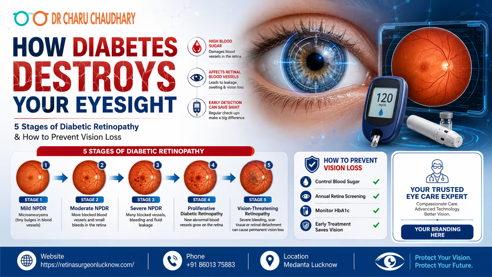
India is currently facing a dual epidemic: the explosion of diabetes and the subsequent rise in vision impairment. As the “Diabetes Capital of the World,” the burden of managing complications like diabetic retinopathy has never been more critical. This condition, often referred to as the “silent thief of sight,” remains the leading cause of preventable blindness in adults worldwide. Diabetic Retinopathy is a diabetes-related eye disease caused by damage to retinal blood vessels. Early diagnosis, good blood sugar control, and regular retina screening can help prevent severe vision loss and blindness. The Hidden Link Between Blood Sugar and Blindness The numbers are startling. According to recent health surveys, nearly one in every three people with diabetes will develop some form of eye damage. In the bustling landscape of Uttar Pradesh, particularly in cities like Lucknow, the prevalence of Type 2 diabetes is shifting toward younger age groups. This shift means people are living with high blood sugar for longer periods, significantly increasing the window for “Diabetic Vision Loss” to occur. Most patients believe that if they can see clearly, their eyes are healthy. This is a dangerous misconception. Diabetic retinopathy often begins without a single symptom. By the time vision becomes blurred or “floaters” appear, the disease has likely progressed to a stage where advanced medical intervention is required. This guide aims to bridge the gap between awareness and action, helping you understand how diabetes affects your eyes and what you can do to stop it. Key Facts About Diabetic Retinopathy To understand the gravity of this condition, let’s look at the data provided by global health leaders like the WHO, International Diabetes Federation (IDF), and the American Academy of Ophthalmology (AAO): What Is Diabetic Retinopathy? At its core, Diabetic Retinopathy is a microvascular complication. The retina is the thin layer of light-sensitive tissue at the back of your eye. It functions like the sensor in a digital camera, capturing light and converting it into electrical signals that the brain interprets as images. For the retina to function, it requires a constant and healthy supply of oxygen and nutrients through a network of tiny, delicate blood vessels. When blood sugar levels remain high for extended periods, it damages the structural integrity of these vessels. They become weak, leak fluid, or close off entirely. This process is the foundation of “Diabetic Eye Disease.” How Diabetes Affects Your Eyesight The destruction of eyesight via diabetes is a progressive, biological cascade: Why Diabetic Eye Damage Often Goes Unnoticed The human brain is remarkably good at compensating for small gaps in vision. In the early stages of retinopathy, the damage usually occurs in the peripheral (side) retina. Because your central vision remains sharp, you may not notice anything is wrong. Furthermore, diabetic eye damage does not cause pain. Unlike a “red eye” or an infection, there is no physical discomfort to alert the patient. This “silent progression” is why annual diabetic eye screening is non-negotiable for every diabetic patient, regardless of their current visual clarity. The 5 Stages of Diabetic Retinopathy Medical professionals categorize the progression of this disease to determine the appropriate treatment path. Stage 1 – Mild Non-Proliferative Diabetic Retinopathy (NPDR) This is the “alert” stage. At this point, tiny, balloon-like swellings called microaneurysms appear in the retinal blood vessels. Stage 2 – Moderate Non-Proliferative Diabetic Retinopathy As the disease advances, more blood vessels swell and lose their ability to transport blood. They may begin to leak blood and fluid, causing the retina to look “spotted” during an exam. Stage 3 – Severe Non-Proliferative Diabetic Retinopathy In this critical stage, a large number of blood vessels are blocked, depriving several areas of the retina of blood flow. These areas secrete growth factors that signal the eye to start growing new vessels. Stage 4 – Proliferative Diabetic Retinopathy (PDR) This is the advanced, vision-threatening stage. The “proliferative” part refers to the rapid growth of new, fragile blood vessels (neovascularization) along the inside surface of the retina and into the vitreous gel. Stage 5 – Advanced Vision-Threatening Diabetic Retinopathy If PDR is left untreated, it leads to severe complications. The abnormal vessels can cause scar tissue to form, which can pull the retina away from the back of the eye (Tractional Retinal Detachment). It can also cause a form of high eye pressure called Neovascular Glaucoma. Stage-by-Stage Risk Chart Stage Pathological Features Vision Risk Recommended Action Stage 1: Mild Microaneurysms Very Low Annual Screening Stage 2: Moderate Vessel leakage & swelling Low 6-Month Monitoring Stage 3: Severe Multiple blocked vessels High 3-Month Monitoring/Laser Stage 4: Proliferative New fragile vessel growth Very High Injections / Laser Stage 5: Advanced Scarring & Detachment Extreme Vitrectomy Surgery Note: While these stages are sequential, a complication called Diabetic Macular Edema (DME) can happen at any of these stages, causing immediate central vision blurriness. Early Symptoms of Diabetic Retinopathy You Should Never Ignore While we emphasize that early stages are silent, you must be on the lookout for these “red flags”: Who Is Most at Risk of Diabetic Vision Loss? Certain factors act as “accelerants” for eye damage: Can Diabetic Retinopathy Cause Permanent Blindness? The short answer is yes, but it is not an inevitability. Blindness occurs when the disease reaches Stage 5, where the retina detaches or the optic nerve is damaged. However, with modern advancements, even patients with advanced stages can often have their vision stabilized. The key distinction is between “preventing” and “restoring.” It is much easier to prevent vision loss than it is to restore it once the retina has been scarred. This highlights the importance of a regular retina checkup. How Doctors Diagnose Diabetic Retinopathy Diagnosing this condition requires more than a simple eye chart test. A retina specialist in Lucknow will use a combination of: Dilated Eye Examination Using drops to enlarge the pupil, the doctor can see the entire retina clearly using a specialized microscope called a slit lamp. Optical Coherence Tomography (OCT Scan) This is a gold-standard diagnostic tool. It’s essentially an “ultrasound with light” that provides high-definition, cross-sectional images of the retina. It can

The human eye is often described as a camera, and the retina is the delicate film at the back that captures every image we see. When the retina is healthy, vision is sharp and vibrant. However, various retinal diseases can cause fluid to leak or abnormal blood vessels to grow, threatening the clarity of our sight. For many years, these conditions led to permanent vision loss, but the advent of Anti-VEGF injections for retina care has revolutionized ophthalmology. While the idea of an “eye injection” can understandably cause anxiety, this procedure is a quick, safe, and highly effective way to preserve and sometimes even improve vision. Anti-VEGF injections for retina diseases help reduce abnormal blood vessel growth and retinal swelling, commonly used in diabetic retinopathy, macular degeneration, and retinal vein occlusion to help protect vision and slow further damage. In this comprehensive guide, we will draw upon the expertise and patient-first philosophy of Dr. Charu Chaudhary, recognized as a trusted Best Retina specialist in Lucknow, to help you understand why these injections are recommended, what the procedure involves, and how they play a vital role in long-term eye health. What Are Anti-VEGF Injections? To understand Anti-VEGF therapy, we first need to understand what VEGF is. VEGF stands for Vascular Endothelial Growth Factor. In a healthy body, this protein is beneficial; it helps form new blood vessels during healing and development. However, in certain retinal diseases, the eye produces too much VEGF. This “over-signaling” causes the growth of weak, abnormal blood vessels that are prone to leaking fluid and blood into the retina. This leakage causes retinal swelling (edema), which distorts vision and can eventually lead to scarring and permanent blindness. Anti-VEGF injections are medications designed to block (or “anti”) this protein. By neutralizing the excess VEGF, the medicine helps: Think of Anti-VEGF as a “leak sealant” for the tiny pipes in your eye. It dries up the excess fluid, allowing the retina to function more normally. Which Retina Diseases May Require Anti-VEGF Injections? Not every eye condition requires injections, but for specific “wet” or “exudative” diseases, they are the gold standard of care. 1. Age-Related Macular Degeneration (Wet AMD) Macular degeneration is a leading cause of vision loss in people over 50. While the “dry” form is more common, the “wet” form is more aggressive. In Wet AMD, abnormal vessels grow under the macula. Without Anti-VEGF injections, these vessels can cause rapid central vision loss, making it difficult to recognize faces or drive. 2. Diabetic Retinopathy and Diabetic Macular Edema (DME) For patients with diabetes, high blood sugar levels can damage the tiny blood vessels in the retina. This leads to diabetic retinopathy. When these damaged vessels leak fluid into the center of the retina, it causes diabetic macular edema. Anti-VEGF injections are incredibly effective at reducing this swelling and preventing the progression of the disease. 3. Retinal Vein Occlusion (RVO) A retinal vein occlusion is essentially a “stroke” in the eye. A blockage in a vein prevents blood from draining out of the retina, causing pressure to build and fluid to leak. This often results in sudden, painless blurring. Anti-VEGF injections help clear the fluid and manage the complications of the blockage. 4. Myopic Choroidal Neovascularization In cases of extreme nearsightedness (high myopia), the retina can become so stretched that it develops cracks, allowing abnormal blood vessels to grow. Anti-VEGF therapy is used here to prevent scarring in the central vision. Common Symptoms That May Indicate Retina Problems Retinal diseases often start quietly. You might not feel any pain, which is why regular eye exams are crucial. However, if you experience any of the following, you should consult a retina specialist immediately: If you notice these signs, Dr. Charu Chaudhary emphasizes that early diagnosis is the key to successful treatment. Waiting too long can allow permanent scarring to occur. How Are Anti-VEGF Injections Given? One of the biggest hurdles for patients is the “fear factor.” It is perfectly natural to feel nervous about a needle near the eye. However, the procedure is far less daunting than most people imagine. A Calm and Controlled Environment Retina injections are performed as an outpatient procedure in a specialized treatment room. The process is designed for maximum safety and minimum discomfort. The entire process, from entering the room to leaving, usually takes about 10 to 15 minutes. Step-by-Step: What Patients Should Expect Before and After Retina Injections Understanding the journey can significantly reduce anxiety. Here is what a typical treatment cycle looks like: Step 1: Retina Examination & Scans Before any injection, you will undergo a comprehensive exam. This usually includes Optical Coherence Tomography (OCT)—a non-invasive scan that provides a cross-sectional view of your retina, showing exactly where the fluid is. Step 2: Eye Preparation On the day of the procedure, your eye will be dilated. The sterile cleaning and numbing process described above will follow. Step 3: The Procedure You will be asked to look in a specific direction while the doctor stabilizes the eye. You won’t see the needle coming toward you, which helps reduce the “flinch” response. Step 4: Short Observation Period You might stay in the clinic for a few minutes. Your doctor may check your eye one last time to ensure there are no immediate issues. Step 5: Temporary Mild Discomfort For the first 24 hours, your eye might feel “gritty” or like there is a piece of sand in it. This is usually due to the antiseptic cleaning solution, not the injection itself. Artificial tears can help. Step 6: Follow-Up Retina Monitoring Anti-VEGF is rarely a “one-and-done” treatment. You will have a follow-up appointment (usually in 4–6 weeks) to see how the retina is responding to the medicine. Step 7: Repeat Injections if Needed Retinal diseases are often chronic. To keep the “pipes from leaking” again, many patients require a series of injections—either monthly or on a “treat-and-extend” schedule where the time between injections is gradually increased. Retina Diseases Commonly Treated with Anti-VEGF (Comparison Chart) Retina Condition Common Symptoms How Anti-VEGF Helps Diabetic Retinopathy Blurry vision, floaters Reduces swelling and prevents vessel growth Wet Macular