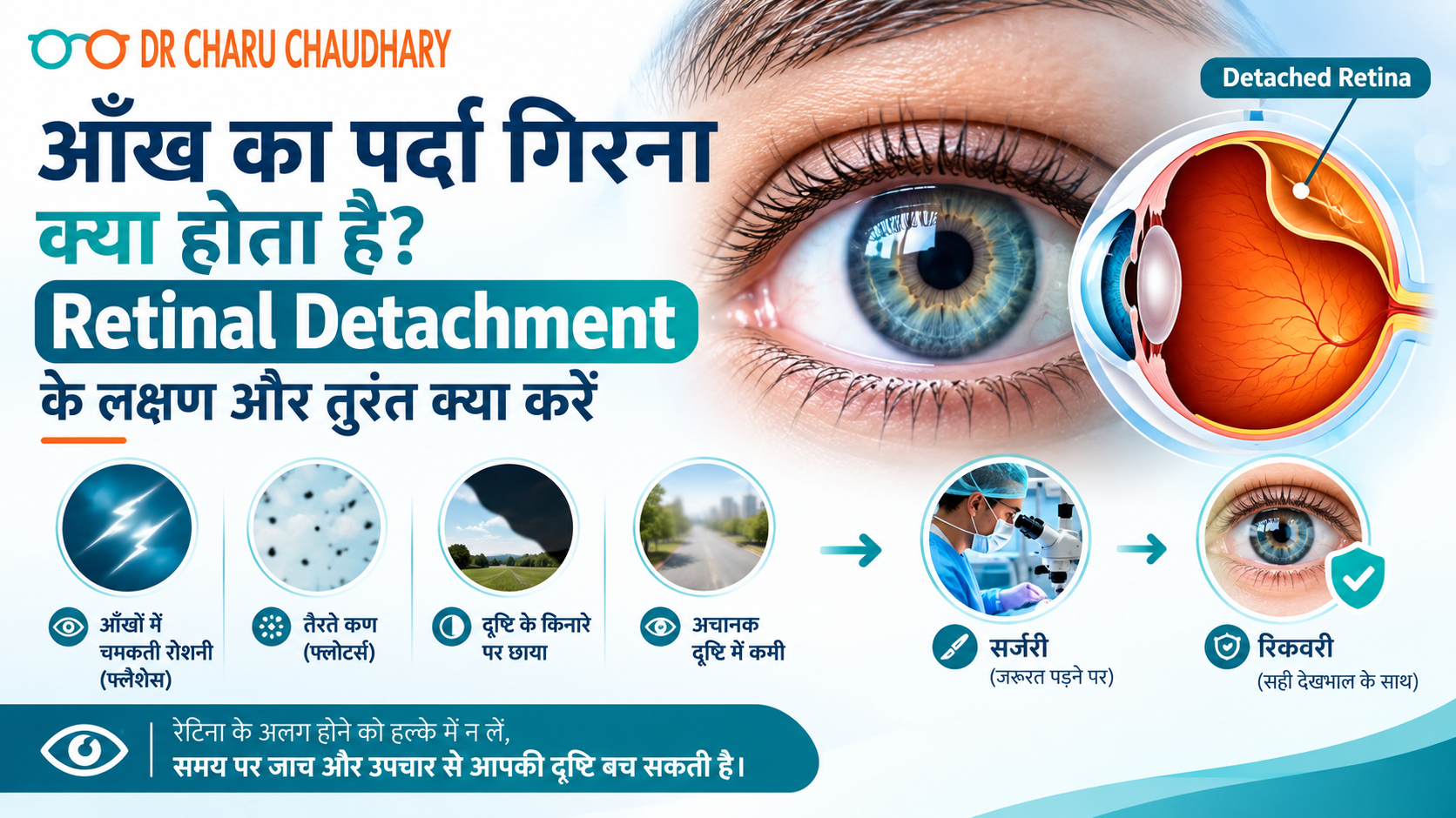
कल्पना कीजिए – आप सुबह उठते हैं और अचानक महसूस होता है कि एक आँख के सामने काला पर्दा गिर रहा है। या शायद रात में आपको बिजली जैसी चमक दिखी और सोचा “थकान होगी।” अगले दिन उसी आँख से धुंधला दिखने लगा। अधिकतर लोग इसे नज़रअंदाज़ कर देते हैं। यही सबसे बड़ी गलती है। जो आप महसूस कर रहे थे, वह Retinal Detachment – यानी आँख का पर्दा गिरना – के शुरुआती संकेत थे। और यह एक ऐसी medical emergency है जिसमें हर घंटा महत्वपूर्ण होता है। मैं Dr. Charu Chaudhary हूँ, Lucknow के Medanta Hospital में Vitreoretinal Surgeon। अपने वर्षों के अनुभव में मैंने देखा है कि जो मरीज़ 24–48 घंटे के भीतर आते हैं, उनकी रोशनी बचाई जा सकती है। जो देर करते हैं – उनके लिए रास्ता बहुत मुश्किल हो जाता है। इस blog में मैं आपको सरल भाषा में बताऊँगी कि आँख का पर्दा गिरना क्या होता है, इसके लक्षण क्या हैं, और सबसे ज़रूरी – इस स्थिति में आपको तुरंत क्या करना चाहिए। रेटिना क्या होती है और वह इतनी ज़रूरी क्यों है? आँख को एक camera की तरह समझिए। जिस तरह camera में film होती है जो तस्वीर कैद करती है – उसी तरह आँख में retina होती है। यह एक पतली, नाज़ुक परत होती है जो आँख के पिछले हिस्से में होती है। जब भी कोई रोशनी आँख में आती है, रेटिना उसे electrical signals में बदलता है और इन signals को Optic Nerve के ज़रिए दिमाग तक पहुँचाता है। दिमाग इन्हें image के रूप में पहचानता है – और आपको दिखता है। अगर यही रेटिना अपनी जगह से हट जाए – तो signal टूट जाता है। दृष्टि धुंधली हो जाती है, गायब हो सकती है, और बिना इलाज के हमेशा के लिए जा सकती है। आँख का पर्दा गिरता क्यों है – मुख्य कारण Retinal Detachment अचानक होता है, लेकिन इसके पीछे कुछ जोखिम कारण होते हैं जिनके बारे में आपको पहले से जानकारी होनी चाहिए: चोट या Trauma: आँख पर तेज़ चोट लगने से, पटाखे फूटने से, या खेल के दौरान चोट लगने से रेटिना खिंच सकती है और अपनी जगह से हट सकती है। High Myopia (तेज़ चश्मा): जिन लोगों का नंबर -6 या उससे अधिक है, उनकी आँख का आकार सामान्य से बड़ा होता है। इससे रेटिना पर खिंचाव रहता है और detachment का खतरा बढ़ जाता है। Diabetes: लम्बे समय से बेकाबू शुगर रेटिना की नसों को कमज़ोर करती है। इसे Diabetic Retinopathy कहते हैं – और यह Retinal Detachment का एक बड़ा कारण बन सकती है। बढ़ती उम्र: उम्र के साथ आँख के अंदर का gel (Vitreous) सिकुड़ता है और रेटिना को खींच सकता है। 50 वर्ष से अधिक उम्र के लोगों में यह ज़्यादा देखा जाता है। पहले का Retinal Detachment: एक आँख में हो चुका है तो दूसरी आँख में भी जोखिम बढ़ जाता है। पारिवारिक इतिहास: परिवार में किसी को हो चुका है तो आपको नियमित जाँच करानी चाहिए। आँख का पर्दा गिरने के लक्षण – जिन्हें कभी नज़रअंदाज़ न करें यह वह हिस्सा है जिसे मैं सबसे ज़्यादा ज़ोर देकर बताना चाहती हूँ। Retinal Detachment में दर्द नहीं होता—यही इसे इतना खतरनाक बनाता है। लोग सोचते हैं “दर्द नहीं तो गंभीर नहीं होगा।” यह सोच गलत है। इन लक्षणों में से कोई भी दिखे – तुरंत doctor के पास जाएँ: अचानक बहुत सारे काले धब्बे या जाले दिखना (Floaters) हाँ, कुछ floaters सामान्य हो सकते हैं। लेकिन अगर अचानक बहुत सारे नए floaters एक साथ दिखें – जैसे मकड़ी का जाला या काली बिंदियाँ तैर रही हों – तो यह रेटिना में खिंचाव या टूटन का संकेत है। बिजली जैसी चमक दिखना (Flashes of Light) आँखें बंद करने पर या अंधेरे में भी चमक दिखे – जैसे camera flash हो। यह इस बात का संकेत है कि Vitreous रेटिना को खींच रहा है। आँख के सामने काला पर्दा या छाया आना ऐसा महसूस हो जैसे किसी ने एक तरफ से curtain खींच दिया हो। पहले side vision जाती है, फिर धीरे-धीरे बीच का हिस्सा भी ढकने लगता है। यह सबसे serious लक्षण है। इस स्थिति में हर मिनट मायने रखता है। अचानक धुंधला दिखना, सिर्फ एक आँख से अचानक एक आँख से कम या धुंधला दिखना – और यह चश्मा लगाने पर भी ठीक न हो। यह इतनी बड़ी Emergency क्यों है? रेटिना एक जीवित tissue है। जब यह अपनी जगह से हट जाती है, तो उसे blood supply मिलनी बंद हो जाती है। जिस तरह खून न मिलने पर कोई भी अंग मरने लगता है – उसी तरह रेटिना की कोशिकाएँ भी मरने लगती हैं। पहले 24 घंटे: अगर पर्दा centre (Macula) तक नहीं पहुँचा, तो surgery के बाद अच्छी रोशनी वापस आने की संभावना बहुत अधिक है। 24–72 घंटे: Macula तक पहुँचने के बाद रोशनी वापस आना मुश्किल होने लगता है। 72 घंटे से अधिक: रेटिना की कोशिकाएँ स्थायी रूप से मर सकती हैं। रोशनी का नुकसान permanent हो सकता है। इसीलिए Retinal Detachment को हम “Golden Hour Emergency” कहते हैं। तुरंत क्या करें – Step by Step अगर आपको या आपके किसी परिवार वाले को ऊपर दिए गए लक्षण दिखें: Step 1: आँख को मलें नहीं, दबाएँ नहीं। Step 2: दोनों आँखें बंद करके शांत रहें – ज़्यादा eye movement से रेटिना खिंच सकता है। Step 3: कोई भी घरेलू उपाय, eye drop, या “देखते हैं कल” – यह सब बंद करें। Step 4: तुरंत किसी Vitreoretinal Specialist से मिलें। General physician या chemist के पास जाने का वक्त नहीं है। Lucknow में हैं तो अभी appointment book करें – हम emergency cases को उसी दिन देखते हैं। Diagnosis कैसे होती है? जब आप clinic आते हैं, तो हम कुछ ज़रूरी जाँचें करते हैं: Dilated Fundus Examination: आँख की पुतली को drops से बड़ा करके रेटिना को सीधे देखा जाता है। B-Scan Ultrasound: अगर आँख के अंदर खून है या साफ नहीं दिख रहा, तो Ultrasound से रेटिना की स्थिति देखी जाती है। OCT (Optical Coherence Tomography): रेटिना की परतों की detailed imaging – यह बताता है कि कितना नुकसान हुआ है। ये सब जाँचें दर्द रहित हैं
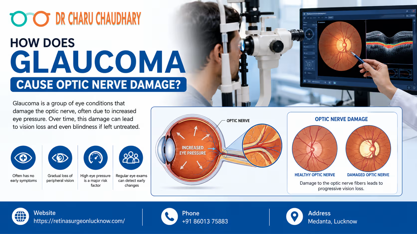
Vision is often considered our most precious sense, yet many of us take it for granted until it begins to fade. Among the various conditions that can threaten our sight, glaucoma stands out as one of the most mysterious and dangerous. Often called the “silent thief of sight,” glaucoma is a group of eye conditions that damage the optic nerve—the vital link between your eye and your brain. Because this damage often happens slowly and without pain, millions of people worldwide are unaware they even have the condition until significant vision loss has occurred. Understanding the relationship between glaucoma and optic nerve health is the first step toward prevention. In this comprehensive guide, we will explore the mechanics of glaucoma optic nerve damage, why early detection is life-changing, and how experts like Dr. Charu Chaudhary are helping patients preserve their vision. How Glaucoma Damages the Optic Nerve Glaucoma causes optic nerve damage by increasing pressure inside the eye or reducing blood supply to the nerve fibers. Over time, this pressure crushes sensitive nerve cells, leading to permanent vision loss if the condition is not detected and treated early. What Is Glaucoma? Understanding the Basics At its core, glaucoma is not just a single disease but a category of ocular disorders characterized by progressive damage to the optic nerve. Understanding Glaucoma in Simple Terms Think of your eye like a sink with a faucet and a drain. The “faucet” produces a clear fluid called aqueous humor to nourish the eye. The “drain” (located at the angle where the iris and cornea meet) allows this fluid to leave. In a healthy eye, the production and drainage are balanced. In glaucoma, the drain gets clogged or works inefficiently, causing fluid to build up. This buildup increases pressure, which eventually pushes against the optic nerve. How Common Is Glaucoma? Glaucoma is a leading cause of irreversible blindness globally. According to the World Health Organization (WHO), it is the second leading cause of blindness after cataracts. However, unlike cataracts, which can be surgically “cured” to restore sight, the vision loss caused by glaucoma is permanent. Why Glaucoma Is a Serious Eye Disease The danger of glaucoma lies in its stealthy nature. In the most common form (open-angle glaucoma), there are no symptoms in the early stages. No pain, no redness, and no sudden blurring. By the time a patient notices a “tunnel vision” effect, up to 40% of the optic nerve fibers may already be destroyed. This is why Dr. Charu Chaudhary emphasizes that regular screenings are the only way to catch the thief before it steals your sight. Can Glaucoma Cause Permanent Blindness? Yes. If left untreated, glaucoma eventually destroys the entire optic nerve, resulting in total blindness. However, with modern medical interventions and early diagnosis by the Best Eye Specialist in Lucknow, the vast majority of patients can maintain functional vision for the rest of their lives. What Is the Optic Nerve and Why Is It Important? To understand glaucoma optic nerve damage, we must first understand what the optic nerve does. How the Optic Nerve Connects the Eye to the Brain The optic nerve is often described as the “electric cable” of the eye. It is composed of more than a million tiny nerve fibers (retinal ganglion cells). These fibers collect visual information from the retina (the light-sensitive tissue at the back of the eye) and transmit it to the brain. How Visual Signals Travel Why Healthy Optic Nerves Are Essential for Vision Without a functional optic nerve, the eye and the brain cannot communicate. Even if your eye is perfectly healthy in every other way—clear lens, healthy retina, perfect cornea—you will be blind if the optic nerve is severed or destroyed. It is the “bridge” of sight. What Happens When the Optic Nerve Gets Damaged? When the fibers within the optic nerve begin to die, the “cable” loses its ability to transmit full images. Initially, the brain compensates for small gaps in the visual field. However, as more fibers die, the gaps become larger, leading to permanent blind spots. How Does Glaucoma Cause Optic Nerve Damage? The process of damage is complex and can involve several biological mechanisms. 1. Increased Eye Pressure (Intraocular Pressure – IOP) High intraocular pressure is the most significant risk factor for glaucoma. When fluid (aqueous humor) cannot drain properly, the pressure inside the eye rises. This pressure exerts physical force on the optic nerve head (the point where the nerve leaves the eye). Over time, this mechanical stress compresses the nerve fibers and the tiny blood vessels that nourish them. 2. Reduced Blood Supply to the Optic Nerve Some patients develop glaucoma even with “normal” eye pressure. This suggests that poor blood flow (ischemia) to the optic nerve also plays a role. If the blood vessels supplying the nerve are narrow or if blood pressure is too low, the nerve cells don’t get enough oxygen and nutrients, leading to cell death. 3. Damage to Retinal Nerve Fibers The optic nerve is made of the axons of retinal ganglion cells. Glaucoma specifically targets these cells. The high pressure or low blood flow triggers a process called “apoptosis” or programmed cell death. Once these cells die, they do not regenerate. 4. Progressive Loss of Nerve Cells The damage usually starts at the outer edges of the optic nerve, which corresponds to our peripheral (side) vision. As the disease progresses, the damage moves inward toward the center, eventually affecting central vision and leading to total blindness. Why the Damage Is Usually Permanent Unlike skin or bone, the nerve cells in the human central nervous system (which includes the optic nerve) do not have the capacity to regrow once they are dead. This is why glaucoma treatment focuses on “saving what’s left” rather than “restoring what’s lost.” The Step-by-Step Process of Glaucoma Damage Glaucoma is a journey of progression. Understanding these stages can help patients realize where they stand. Stage What is Happening? Impact on Vision Stage 1: Increased Pressure Fluid drainage slows down; IOP begins to rise. No noticeable
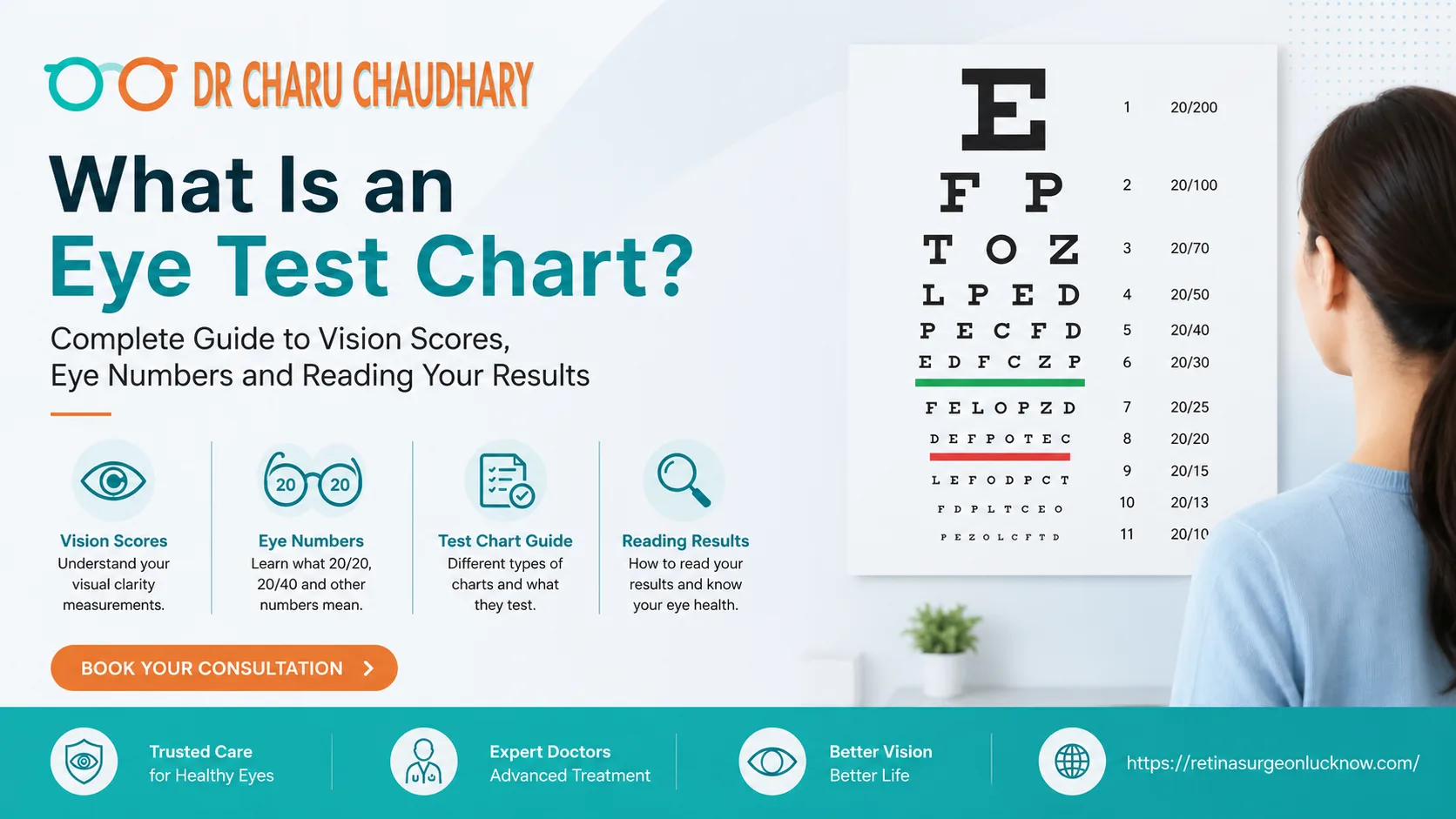
An eye test chart, most commonly the Snellen chart, is a clinical tool used by eye specialists to measure visual acuity and sharpness. During an eye exam, patients read rows of letters or symbols from a specific distance to determine their vision score, such as 20/20 or 6/6. These charts help doctors identify vision errors and determine if glasses or further medical treatments are necessary. Have you ever sat in a dim exam room, covering one eye, and trying to read a line of letters that seem to shrink as they go down? Most of us have encountered the classic “E” chart at some point. As an eye specialist, I, Dr. Charu Chaudhary, find that while many people have taken this test, very few actually understand what the results mean. “Does a minus number mean I’m going blind?” or “Is 20/20 vision perfect health?” are questions I hear daily. Understanding your vision shouldn’t be a mystery. This guide is designed to simplify the Snellen chart, decode those confusing eye numbers, and help you interpret your eye reports with confidence. Quick Summary If you are looking for a fast overview of vision scores, here are the essentials: What Is an Eye Test Chart? At its core, an eye test chart is a standardized way to measure “visual acuity.” Visual acuity refers to the sharpness or clarity of your vision at a specific distance. When you visit a clinic, we use these charts to determine if your eyesight meets the “normal” standard or if you have a refractive error that requires correction. The chart consists of various rows of “optotypes”—specially designed letters or symbols. While the chart looks simple, it is a scientifically calibrated tool. Each row represents a different level of visual ability. If you can read the small lines at the bottom, your brain and eyes are working together efficiently to process fine detail. The Snellen Chart — The Gold Standard of Vision Testing The most recognizable eye test chart in the world is the Snellen Chart. Developed in 1862 by Dutch ophthalmologist Herman Snellen, it remains the primary tool used by eye specialists like myself. How the Snellen Chart Works The chart typically features a large letter “E” at the top, followed by rows of letters that get progressively smaller. In my practice, I use the Snellen chart not just to prescribe glasses, but as a “vital sign” for the eye. A sudden drop in your ability to read the Snellen lines can alert us to underlying issues like cataracts or retinal changes. 6 Types of Eye Test Charts — Which One Is Right for You? While the Snellen chart is the most famous, eye specialists use various charts depending on the patient’s age and specific needs. 1. The Classic Snellen Chart Used for adults and children who know the alphabet. It uses a specific set of 10 letters (C, D, E, F, L, N, O, P, T, Z) known as Snellen optotypes. 2. The Tumbling E Chart This is used for people who cannot read the alphabet or for young children. The patient simply indicates which direction the “fingers” of the letter E are pointing (up, down, left, or right). 3. Landolt C Chart Similar to the Tumbling E, this chart uses a circle with a gap (like the letter C). The patient identifies where the gap is located. It is often used in international research for its high level of accuracy. 4. Pediatric Charts (LEA Symbols) For very young children, we use symbols like houses, apples, and hearts. This allows us to test a child’s vision before they even learn their ABCs. 5. Near Vision Charts (Jaeger Chart) This is a small, hand-held card used to test how well you see up close. It is essential for diagnosing “Presbyopia,” the age-related loss of near-focusing ability that usually begins after age 40. 6. Pelli-Robson Contrast Sensitivity Chart This chart doesn’t just measure size; it measures how well you can see objects against a background. This is crucial for patients with glaucoma or those who struggle with night driving. 📊Vision Score Chart — What Is a Normal Eye Test Reading? What does your score actually say about your eyesight? Here is a simple breakdown of common Snellen chart readings. Vision Score (Feet) Vision Score (Metric) Meaning 20/20 6/6 Normal Vision: You see at 20ft what a normal person sees at 20ft. 20/15 6/4.5 Excellent Vision: You see better than the average person. 20/40 6/12 Mild Blur: Most states require at least this for a driver’s license. 20/70 6/21 Moderate Impairment: You may struggle to read standard print without help. 20/200 6/60 Severe Impairment: This is the threshold for legal blindness in many regions. What is a “good” eye test score? A score of 20/20 is the clinical standard for “good” vision. However, having 20/20 vision does not necessarily mean your eyes are perfectly healthy—it just means they are seeing clearly at a distance. What is a “bad” eyesight score? If your score is 20/50 or higher (the second number is larger), your vision is significantly limited. At this stage, everyday tasks like recognizing faces across a street or reading street signs become difficult. 📊How to Read Your Eye Test Results — Prescription Decoder After you finish reading the Snellen chart, your specialist will give you a prescription. These numbers can be confusing. Let’s decode them into patient-friendly language. Term What it Means Simple Explanation OD Oculus Dexter Your Right Eye. OS Oculus Sinister Your Left Eye. SPH (Sphere) The main power Minus (-) means Nearsighted; Plus (+) means Farsighted. CYL (Cylinder) Astigmatism This corrects the “football shape” of your cornea. Axis The Angle The direction (0–180°) the astigmatism correction is placed. ADD Reading Power The “extra” boost needed for reading (bifocals/progressives). How do I read my eye test results? If your report says OD: -2.00 SPH, it means your right eye is nearsighted and needs two diopters of correction to see the 20/20 line on the Snellen chart. If it says +1.50 SPH, you are farsighted, and your
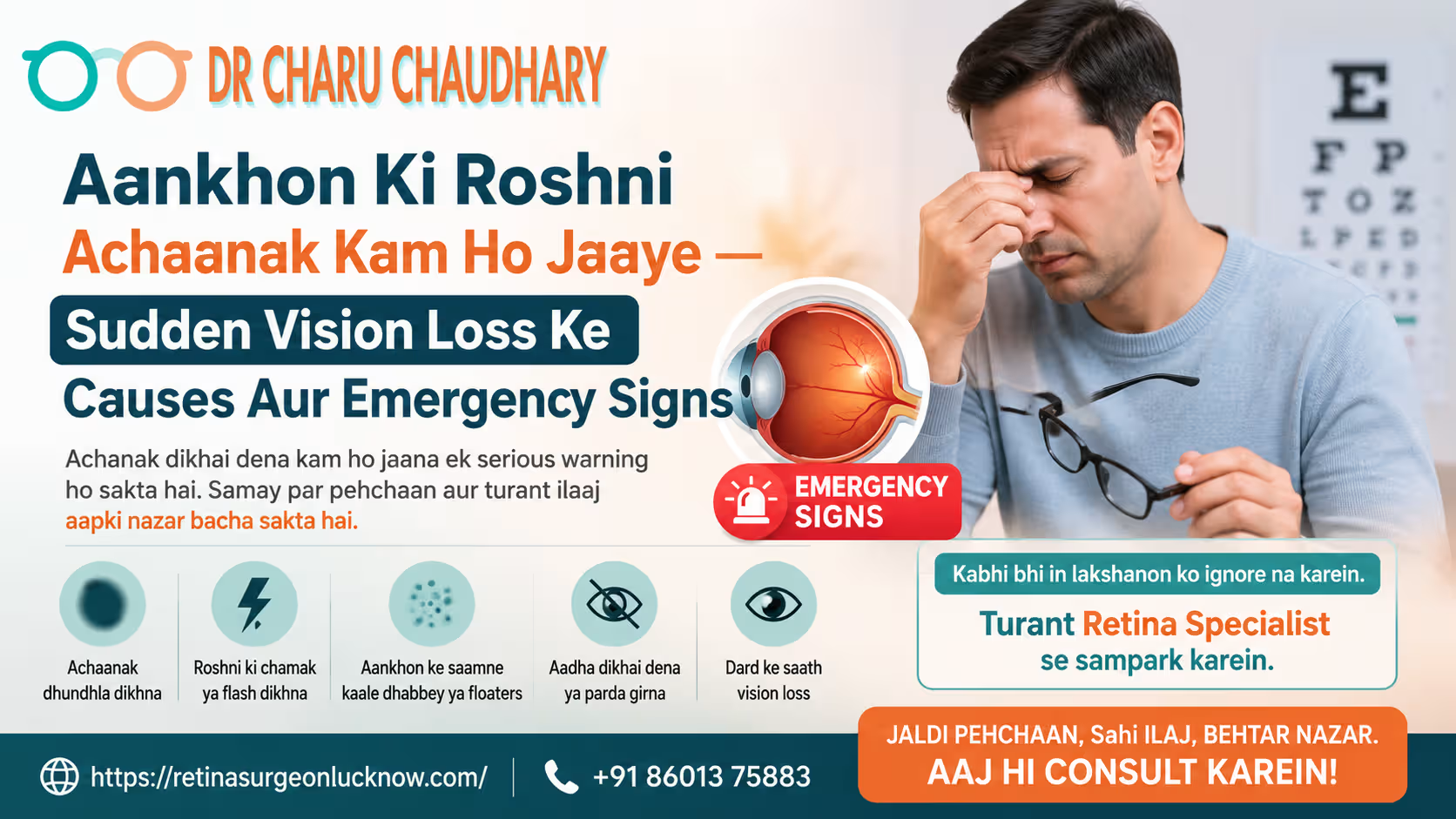
आंखों की रोशनी अचानक कम हो जाए: Sudden Vision Loss के मुख्य कारण, लक्षण और Emergency Signs आंखें हमारे शरीर का सबसे संवेदनशील हिस्सा हैं। यदि अचानक आपको धुंधला दिखने लगे या एक आंख से रोशनी पूरी तरह चली जाए, तो यह स्थिति किसी भी व्यक्ति को डरा सकती है। मेडिकल भाषा में इसे Sudden Vision Loss कहा जाता है। अक्सर लोग इसे कमजोरी या थकान समझकर टाल देते हैं, लेकिन यह एक गंभीर Retina Emergency हो सकती है। एक अनुभवी Retina specialist in Lucknow होने के नाते, मैं (Dr. Charu Chaudhary) हमेशा मरीजों को सलाह देती हूँ कि आंखों में अचानक होने वाला कोई भी बदलाव “Golden Hour” के भीतर डॉक्टर तक पहुंचना चाहिए। इस ब्लॉग में हम विस्तार से जानेंगे कि अचानक रोशनी जाने के कारण क्या हैं और आपको कब तुरंत डॉक्टर के पास भागना चाहिए। Sudden Vision Loss Kya Hota Hai? Sudden vision loss का मतलब है कुछ ही सेकंड, मिनट या घंटों के भीतर आपकी देखने की क्षमता का कम हो जाना या पूरी तरह खत्म हो जाना। यह स्थिति अलग-अलग लोगों में अलग तरह से दिख सकती है: यह समझना जरूरी है कि अचानक रोशनी जाना कोई सामान्य बात नहीं है। यह आंखों के पीछे के पर्दे (Retina) या दिमाग तक सिग्नल ले जाने वाली नस (Optic Nerve) में किसी गंभीर खराबी का संकेत है। Symptoms Jo Ignore Nahi Karne Chahiye (Emergency Checklist) अगर आपको अपनी आंखों में नीचे दिए गए लक्षणों में से कुछ भी महसूस हो रहा है, तो समझ लें कि यह एक इमरजेंसी है: इन लक्षणों का मतलब हो सकता है कि आपके रेटिना में कोई गंभीर समस्या है। ऐसी स्थिति में Best eye specialist in Lucknow से तुरंत संपर्क करना जीवन भर के अंधेपन से बचा सकता है। Sudden Vision Loss Ke Common Causes अचानक रोशनी कम होने के पीछे कई चिकित्सीय कारण हो सकते हैं। आइए इन्हें विस्तार से समझते हैं: 1. Retina Detachment (परदे का फटना) रेटिना आंख का वह हिस्सा है जो इमेज बनाता है। अगर यह अपनी जगह से खिसक जाए या फट जाए, तो रोशनी अचानक जा सकती है। यह एक बड़ी रेटिना इमरजेंसी है। 2. Eye Stroke (Vascular Occlusion) जैसे दिमाग में स्ट्रोक होता है, वैसे ही आंखों की नसों में खून का थक्का जम सकता है। इसे Sudden loss of vision in one eye का प्रमुख कारण माना जाता है। इसमें दर्द नहीं होता, लेकिन रोशनी तुरंत चली जाती है। 3. Optic Neuritis (नस में सूजन) आंखों की नस (Optic Nerve) में सूजन आने से दिमाग तक सिग्नल नहीं पहुंच पाते। यह मल्टीपल स्क्लेरोसिस जैसी बीमारियों का शुरुआती लक्षण हो सकता है। 4. Acute Glaucoma (काला मोतिया का अटैक) जब आंख के अंदर का दबाव (IOP) अचानक बढ़ जाता है, तो रोशनी कम हो जाती है और आंखों में बहुत तेज दर्द होता है। 5. Migraine Aura कभी-कभी माइग्रेन के कारण कुछ सेकंड या मिनटों के लिए रोशनी जा सकती है। इसे temporary blindness in both eyes के रूप में देखा जा सकता है, जो बाद में ठीक हो जाता है। 6. Vitamin Deficiencies Loss of vision is caused by a deficiency of Vitamin A और Vitamin B12। हालांकि यह अचानक नहीं होता, लेकिन लंबे समय तक कमी रहने पर नसों की कमजोरी अचानक विजन लॉस का कारण बन सकती है। Temporary vs Serious Vision Loss (Quick Comparison Chart) परिस्थिति (Condition) अस्थाई (Temporary) इमरजेंसी (Emergency) मुख्य लक्षण Migraine Aura ✔ ✘ 20-30 मिनट में विजन ठीक हो जाना Retina Detachment ✘ ✔ आंखों के सामने पर्दा आना, फ्लैश दिखना Eye Stroke ✘ ✔ बिना दर्द के अचानक रोशनी जाना Dry Eyes ✔ ✘ बार-बार पलक झपकाने पर साफ दिखना Acute Glaucoma ✘ ✔ तेज दर्द, लाली और धुंधलापन नोट: यदि आप सुनिश्चित नहीं हैं, तो हमेशा डॉक्टर की सलाह लें। देर करने से बेहतर है कि जांच करा ली जाए। Retina Emergency Ke Warning Signs एक Retina specialist in Lucknow होने के नाते, मैं अक्सर देखती हूँ कि मरीज ‘Floaters’ या ‘Flashes’ को नज़रअंदाज कर देते हैं। ये रेटिना फटने के शुरुआती संकेत हैं। यदि आपको लखनऊ या आसपास के क्षेत्रों में ये लक्षण महसूस हों, तो तुरंत विशेषज्ञ की मदद लें। Diabetes Aur High BP Ka Connection जिन लोगों को डायबिटीज (Sugar) या हाई ब्लड प्रेशर है, उन्हें विजन लॉस का खतरा सबसे ज्यादा होता है। डायबिटीज के मरीजों को साल में कम से कम दो बार अपने रेटिना की जांच जरूर करानी चाहिए। Kab Turant Eye Doctor Ke Paas Jaana Chahiye? अचानक विजन लॉस के मामले में “इंतजार करना” सबसे बड़ी गलती है। आपको तुरंत डॉक्टर के पास जाना चाहिए यदि: डॉ. चारु चौधरी जैसे विशेषज्ञों द्वारा Immediate evaluation आपको स्थायी अंधेपन से बचा सकता है। Treatment Delay Karne Ka Risk अगर अचानक रोशनी जाने पर आप 24 से 48 घंटों के भीतर इलाज नहीं कराते, तो: Sudden Vision Loss Ka Treatment Kaise Kiya Jaata Hai? आज के समय में मेडिकल साइंस ने बहुत तरक्की कर ली है। इलाज इस बात पर निर्भर करता है कि कारण क्या है: FAQs Section (Sudden Vision Loss) Q1: What causes sudden loss of vision in one eye?A: इसके मुख्य कारण Eye Stroke, Retina Detachment, या Optic Nerve में सूजन हो सकते हैं। Q2: Sudden loss of vision in one eye treatment kya hai?A: इसका इलाज कारण पर निर्भर करता है, जिसमें लेज़र, सर्जरी या इंजेक्शन शामिल हो सकते हैं। Q3: Sudden painful loss of vision causes kya ho sakte hain?A: दर्द के साथ रोशनी जाना Acute Glaucoma, आंखों में इन्फेक्शन (Uveitis) या गंभीर चोट का संकेत है। Q4: What can cause temporary blindness in both eyes?A: यह Migraine Aura, लो ब्लड प्रेशर, या मस्तिष्क में खून की कमी के कारण हो सकता है। Q5: Retina emergency ke symptoms kya hote hain?A: अचानक फ्लैश दिखना, बहुत सारे फ्लोटर्स (Floaters) और आंखों के सामने काला पर्दा आना। Q6: Kya sudden vision loss permanent ho sakta hai?A: हाँ, अगर सही समय पर इलाज न मिले, तो यह स्थायी अंधापन बन सकता है। 🧑⚕️ Dr. Charu Chaudhary’s Expert Advice एक रेटिना विशेषज्ञ के रूप में, मैंने कई ऐसे केस देखे हैं जहाँ मरीजों ने “कल देखेंगे” सोचकर अपनी रोशनी खो दी। अचानक विजन लॉस एक ऐसी स्थिति है जहाँ हर मिनट कीमती है। खासकर
Retinopathy of Prematurity (ROP) is an eye condition that affects premature babies when abnormal blood vessels grow in the retina. If not detected early, ROP can lead to vision problems or even blindness. Timely ROP screening and treatment are extremely important to protect a baby’s eyesight and ensure healthy development. Bringing a premature baby into the world is a journey filled with both hope and anxiety. As a parent, you are likely navigating a whirlwind of medical terms, NICU monitors, and specialized care plans. Among these concerns, one of the most critical—yet often misunderstood—is Retinopathy of Prematurity (ROP). It is a condition that specifically targets the delicate eyes of our littlest fighters. As a retina specialist, I have sat with many parents, holding their hands as we discuss their baby’s eye health. My goal today is to help you understand ROP, why screening is vital, and how we can work together to protect your baby’s precious sight. What Is Retinopathy of Prematurity (ROP)? To understand ROP, think of the eye like a high-tech camera. The retina is the “film” at the back of the camera that captures images and sends them to the brain. In a full-term baby, the blood vessels that nourish the retina finish growing just before birth. However, when a baby is born too early, these vessels haven’t reached the edges of the retina yet. After birth, these vessels can start growing in a disorganized or “abnormal” way. Instead of lying flat, they might leak or bleed, leading to scarring. If this scarring pulls the retina away from the back of the eye, it causes vision loss. Premature babies are at risk because their internal systems are still learning to adapt to the world outside the womb. This is why consulting a Retina specialist in Lucknow for early evaluation is a non-negotiable step in your baby’s NICU journey. Which Babies Are at Higher Risk for ROP? Not every premature baby will develop ROP, but certain factors increase the risk significantly. We closely monitor babies who meet the following criteria: If your baby falls into these categories, please do not panic. Most babies with ROP have mild cases that resolve on their own, but we must watch them like a hawk to be sure. Retinopathy of Prematurity Symptoms Parents Should Know One of the most challenging things about ROP is that it has no visible symptoms in its early stages. You cannot see ROP by looking at your baby’s eyes in the nursery. There is no redness, no tearing, and no obvious pain. This is why the ROP eye test for premature babies is the only way to know what is happening. However, as a child grows, some signs might suggest that ROP occurred or is affecting their vision: Because early ROP is “invisible,” we rely entirely on clinical screening to save sight. 📊 Retinopathy of Prematurity Stages (Quick Chart) We categorize ROP into stages based on the severity of the abnormal vessel growth. ROP Stage What It Means Stage 1 Mildly abnormal blood vessel growth. Most babies get better without treatment. Stage 2 Moderate abnormal growth. Usually improves on its own but requires close monitoring. Stage 3 Severe abnormal growth. Vessels become tangled and scarred. Treatment is often needed. Stage 4 Partial retinal detachment. The retina begins to pull away from the eye wall. Stage 5 Complete retinal detachment. This is the most severe stage and can lead to blindness. Understanding the Stages: Stages 1 and 2 often resolve naturally as the baby grows. However, when a baby reaches Stage 3 “plus disease” (where vessels become dilated and twisted), we move quickly toward treatment. ROP stage 5 treatment is much more complex and involves major surgery, which is why we aim to catch everything at Stage 3 or earlier. Why Is the ROP Eye Test Important for Premature Babies? The window of opportunity to treat ROP is very small. If we miss the “threshold” for treatment, the condition can progress to permanent blindness within days. Timely screening is the only way to: I often tell parents in my clinic that “timing is everything.” Parents are strongly advised to consult a trusted eye specialist in Lucknow as soon as the neonatologist recommends an eye check, usually within 3 to 4 weeks after birth. How Is the ROP Screening Procedure Done? It is natural to feel protective when you see your tiny baby undergoing tests. Rest assured, the ROP procedure is a standard, safe, and quick examination. The exam usually takes only a few minutes. Your baby might cry a little because they don’t like being held still, but they are not in significant pain. ROP in Premature Babies — Treatment Options If we find that the ROP is progressing to a stage that threatens vision, we have several effective tools. Retinopathy of Prematurity Treatment in Lucknow has advanced significantly, offering babies a great chance at normal vision. Dr. Charu Chaudhary is known for advanced retina care and timely ROP management, ensuring that every baby receives a customized treatment plan based on their specific stage. A Story of Hope: The Journey of Baby Kabir I remember Baby Kabir, born at just 28 weeks. His parents were terrified when I told them he had reached Stage 3 ROP. They felt like they had already been through enough in the NICU. We performed a laser procedure the very next day. It was a stressful hour for the parents, but the procedure was a success. Today, Kabir is three years old. He wears glasses for mild nearsightedness, but he can see the world, chase his dog, and read his favorite picture books. Early screening was the hero of Kabir’s story. It can be the hero of your baby’s story, too. Recovery and Long-Term Vision Care After ROP Treatment Treatment is the first step, but the journey doesn’t end there. Even after successful treatment, premature babies need long-term follow-ups. Regular visits to a pediatric retina specialist ensure that any minor issues are caught before they affect your child’s learning and development. How Parents Can Protect Their