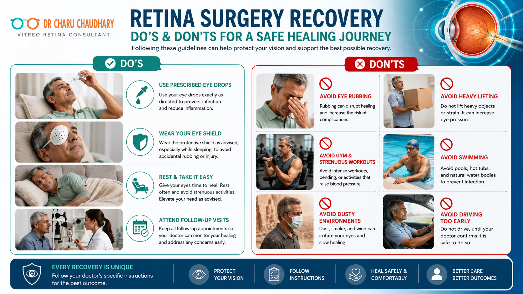
The success of a retinal procedure does not end when the surgeon steps out of the operating room. In fact, for many patients, the most critical phase begins the moment they head home. The retina is a delicate, light-sensitive tissue at the back of the eye, responsible for translating light into the images we see. Because it is so fragile, surgery involving the retina—whether to repair a detachment, clear a hemorrhage, or fix a macular hole—requires a meticulous and disciplined recovery process. Understanding the do’s and don’ts after retina surgery is essential for protecting your vision and ensuring the surgical site heals correctly. Many patients feel anxious about what they can and cannot do. This guide is designed to provide comprehensive, expert-backed information to help you navigate your recovery journey with confidence. Recovery after retina surgery requires careful eye protection, proper medication use, follow-up appointments, and activity restrictions. Following your surgeon’s instructions can reduce complications, support healing, and improve long-term visual outcomes. Understanding Retina Surgery What Is the Retina? The retina is a thin layer of neural tissue lining the inner back surface of the eye. Think of it as the “film” in a traditional camera. It captures light rays that enter the eye and converts them into electrical impulses that the brain interprets as images. If the retina is damaged, scarred, or detached, the “film” becomes distorted or blank, leading to significant vision loss or even permanent blindness. Common Conditions Requiring Retina Surgery Retinal surgery is usually recommended when conservative treatments are insufficient. Common conditions include: Types of Retina Surgery Modern ophthalmology utilizes advanced techniques to repair these issues. According to Dr Charu Chaudhary, a renowned expert and the Best Retina Specialist in Lucknow, understanding your specific procedure helps in adhering to recovery protocols. Vitrectomy This is the most common retina surgery. The surgeon removes the vitreous gel (the clear fluid filling the eye) to better access the retina. The vitreous is then replaced with a saline solution, a gas bubble, or silicone oil. Retinal Detachment Surgery Techniques include Scleral Buckling (placing a flexible band around the eye to push the wall against the retina) or Pneumatic Retinopexy (injecting a gas bubble into the eye to push the retina back into place). Macular Hole Surgery Usually involves a vitrectomy followed by “peeling” a very thin membrane from the surface of the retina to encourage the hole to close. A gas bubble is almost always used here. Epiretinal Membrane Surgery Similar to macular hole surgery, the surgeon removes the vitreous and then delicately peels the scar tissue (membrane) off the retina to reduce distortion. What to Expect Immediately After Retina Surgery The first few hours and days following surgery are often the most uncomfortable, but they are also the most vital for long-term success. First 24 Hours Immediately after surgery, you will likely wear an eye patch and a protective plastic shield. You may feel groggy from sedation. It is normal to feel a “scratchy” sensation, as if there is sand in your eye. This is often due to the tiny incisions or sutures used during the procedure. Vision Changes After Surgery Do not be alarmed if your vision is extremely blurry or if you can only see light and shadows immediately after surgery. If a gas bubble was used, your vision will be blocked by the bubble, making it feel like you are looking through water or a dark curve. As the bubble dissipates, your vision will gradually clear from the top down. Eye Discomfort and Redness The white part of your eye (the sclera) may appear very red or even bloodshot. This is a common side effect of the surgical manipulation and will resolve over 2–3 weeks. Mild aching is normal and can usually be managed with over-the-counter pain relief recommended by your specialist. Protective Eye Shield You will be instructed to wear a protective shield, especially while sleeping, for at least the first week. This prevents accidental rubbing or pressure on the eye during the night. Recovery Timeline After Retina Surgery Recovery is a marathon, not a sprint. Below is a general timeline for recovery after retina surgery. Recovery Period What Patients Can Expect First 24 Hours Patching of the eye, significant blurring, mild pain, and the need for total rest. First Week Frequent use of antibiotic/steroid eye drops; strict head positioning (if a bubble was used); restricted activity. 2–4 Weeks Redness fades; vision begins to stabilize; gas bubble (if used) starts to shrink; can often return to light office work. 1–3 Months Most activity restrictions are lifted; vision continues to improve; final eye glass prescription may be updated. 3–6 Months Full healing achieved; the “new normal” for vision is established; long-term monitoring continues. Note: Every patient heals differently. Always follow the specific timeline provided by Dr Charu Chaudhary or your attending retina specialist. Important Do’s After Retina Surgery Use Eye Drops Exactly as Prescribed Your surgeon will prescribe a combination of antibiotic drops (to prevent infection) and steroid drops (to reduce inflammation). Attend All Follow-Up Visits Post-operative appointments are non-negotiable. Your surgeon needs to monitor the intraocular pressure (IOP) and ensure the retina is staying in place. Missing an appointment could mean missing early signs of a complication. Maintain Proper Head Positioning If a gas or oil bubble was placed in your eye, you may be required to maintain a specific head position (face-down or side-lying) for 23 hours a day for 1–2 weeks. This ensures the bubble floats to the correct spot to “plug” the retinal tear or hole. Protect Your Eye From Injury Wear your eye shield as instructed. Even during the day, wearing your regular glasses can provide a physical barrier against accidental pokes or dust. Get Adequate Rest Your body heals faster when it is well-rested. Avoid the temptation to “be productive” during the first week. Focus on sleeping and staying relaxed. Keep Blood Sugar and BP Under Control High blood sugar or blood pressure can interfere with the healing of delicate retinal blood vessels. This is especially
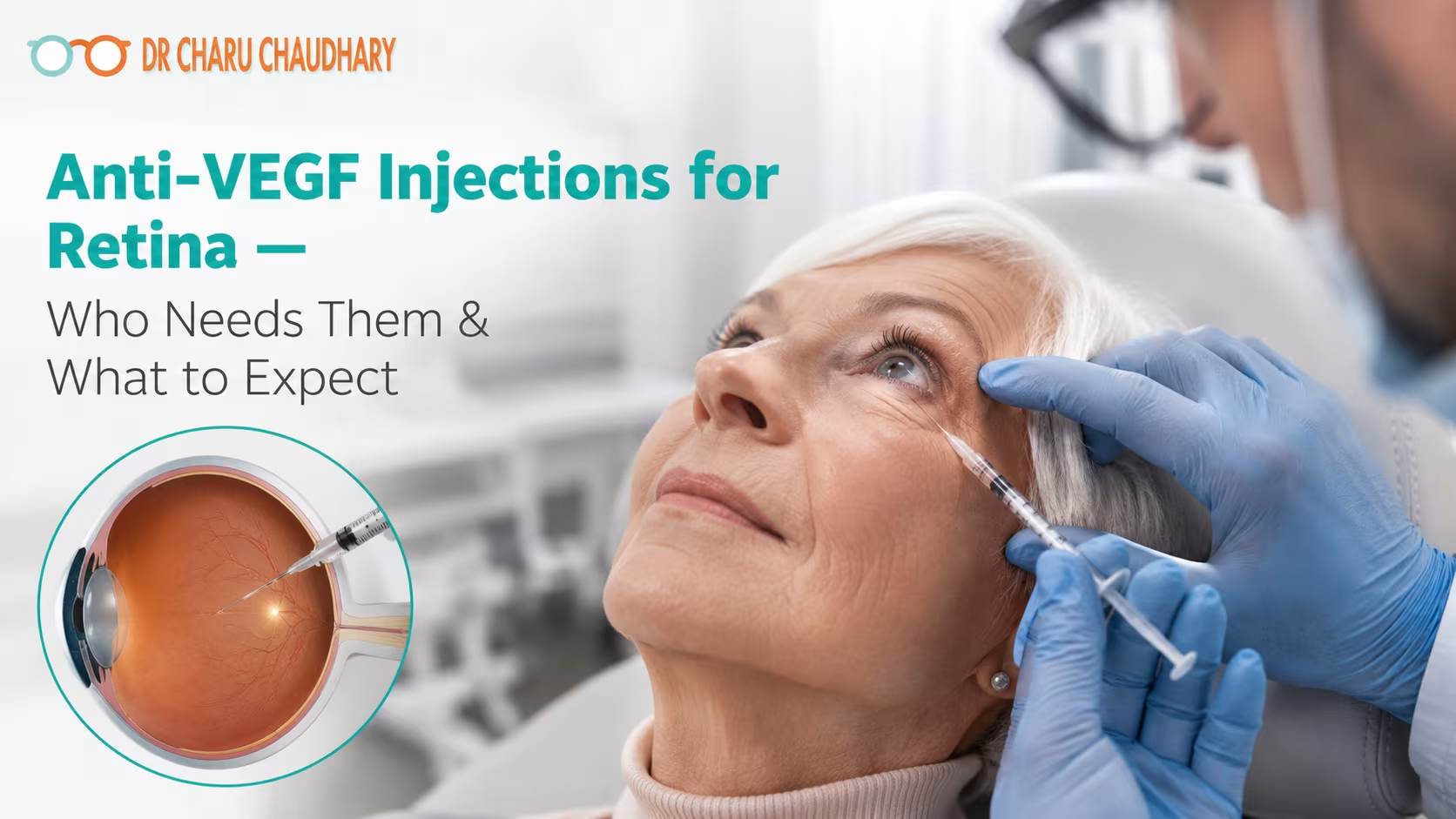
The human eye is often described as a camera, and the retina is the delicate film at the back that captures every image we see. When the retina is healthy, vision is sharp and vibrant. However, various retinal diseases can cause fluid to leak or abnormal blood vessels to grow, threatening the clarity of our sight. For many years, these conditions led to permanent vision loss, but the advent of Anti-VEGF injections for retina care has revolutionized ophthalmology. While the idea of an “eye injection” can understandably cause anxiety, this procedure is a quick, safe, and highly effective way to preserve and sometimes even improve vision. Anti-VEGF injections for retina diseases help reduce abnormal blood vessel growth and retinal swelling, commonly used in diabetic retinopathy, macular degeneration, and retinal vein occlusion to help protect vision and slow further damage. In this comprehensive guide, we will draw upon the expertise and patient-first philosophy of Dr. Charu Chaudhary, recognized as a trusted Best Retina specialist in Lucknow, to help you understand why these injections are recommended, what the procedure involves, and how they play a vital role in long-term eye health. What Are Anti-VEGF Injections? To understand Anti-VEGF therapy, we first need to understand what VEGF is. VEGF stands for Vascular Endothelial Growth Factor. In a healthy body, this protein is beneficial; it helps form new blood vessels during healing and development. However, in certain retinal diseases, the eye produces too much VEGF. This “over-signaling” causes the growth of weak, abnormal blood vessels that are prone to leaking fluid and blood into the retina. This leakage causes retinal swelling (edema), which distorts vision and can eventually lead to scarring and permanent blindness. Anti-VEGF injections are medications designed to block (or “anti”) this protein. By neutralizing the excess VEGF, the medicine helps: Think of Anti-VEGF as a “leak sealant” for the tiny pipes in your eye. It dries up the excess fluid, allowing the retina to function more normally. Which Retina Diseases May Require Anti-VEGF Injections? Not every eye condition requires injections, but for specific “wet” or “exudative” diseases, they are the gold standard of care. 1. Age-Related Macular Degeneration (Wet AMD) Macular degeneration is a leading cause of vision loss in people over 50. While the “dry” form is more common, the “wet” form is more aggressive. In Wet AMD, abnormal vessels grow under the macula. Without Anti-VEGF injections, these vessels can cause rapid central vision loss, making it difficult to recognize faces or drive. 2. Diabetic Retinopathy and Diabetic Macular Edema (DME) For patients with diabetes, high blood sugar levels can damage the tiny blood vessels in the retina. This leads to diabetic retinopathy. When these damaged vessels leak fluid into the center of the retina, it causes diabetic macular edema. Anti-VEGF injections are incredibly effective at reducing this swelling and preventing the progression of the disease. 3. Retinal Vein Occlusion (RVO) A retinal vein occlusion is essentially a “stroke” in the eye. A blockage in a vein prevents blood from draining out of the retina, causing pressure to build and fluid to leak. This often results in sudden, painless blurring. Anti-VEGF injections help clear the fluid and manage the complications of the blockage. 4. Myopic Choroidal Neovascularization In cases of extreme nearsightedness (high myopia), the retina can become so stretched that it develops cracks, allowing abnormal blood vessels to grow. Anti-VEGF therapy is used here to prevent scarring in the central vision. Common Symptoms That May Indicate Retina Problems Retinal diseases often start quietly. You might not feel any pain, which is why regular eye exams are crucial. However, if you experience any of the following, you should consult a retina specialist immediately: If you notice these signs, Dr. Charu Chaudhary emphasizes that early diagnosis is the key to successful treatment. Waiting too long can allow permanent scarring to occur. How Are Anti-VEGF Injections Given? One of the biggest hurdles for patients is the “fear factor.” It is perfectly natural to feel nervous about a needle near the eye. However, the procedure is far less daunting than most people imagine. A Calm and Controlled Environment Retina injections are performed as an outpatient procedure in a specialized treatment room. The process is designed for maximum safety and minimum discomfort. The entire process, from entering the room to leaving, usually takes about 10 to 15 minutes. Step-by-Step: What Patients Should Expect Before and After Retina Injections Understanding the journey can significantly reduce anxiety. Here is what a typical treatment cycle looks like: Step 1: Retina Examination & Scans Before any injection, you will undergo a comprehensive exam. This usually includes Optical Coherence Tomography (OCT)—a non-invasive scan that provides a cross-sectional view of your retina, showing exactly where the fluid is. Step 2: Eye Preparation On the day of the procedure, your eye will be dilated. The sterile cleaning and numbing process described above will follow. Step 3: The Procedure You will be asked to look in a specific direction while the doctor stabilizes the eye. You won’t see the needle coming toward you, which helps reduce the “flinch” response. Step 4: Short Observation Period You might stay in the clinic for a few minutes. Your doctor may check your eye one last time to ensure there are no immediate issues. Step 5: Temporary Mild Discomfort For the first 24 hours, your eye might feel “gritty” or like there is a piece of sand in it. This is usually due to the antiseptic cleaning solution, not the injection itself. Artificial tears can help. Step 6: Follow-Up Retina Monitoring Anti-VEGF is rarely a “one-and-done” treatment. You will have a follow-up appointment (usually in 4–6 weeks) to see how the retina is responding to the medicine. Step 7: Repeat Injections if Needed Retinal diseases are often chronic. To keep the “pipes from leaking” again, many patients require a series of injections—either monthly or on a “treat-and-extend” schedule where the time between injections is gradually increased. Retina Diseases Commonly Treated with Anti-VEGF (Comparison Chart) Retina Condition Common Symptoms How Anti-VEGF Helps Diabetic Retinopathy Blurry vision, floaters Reduces swelling and prevents vessel growth Wet Macular