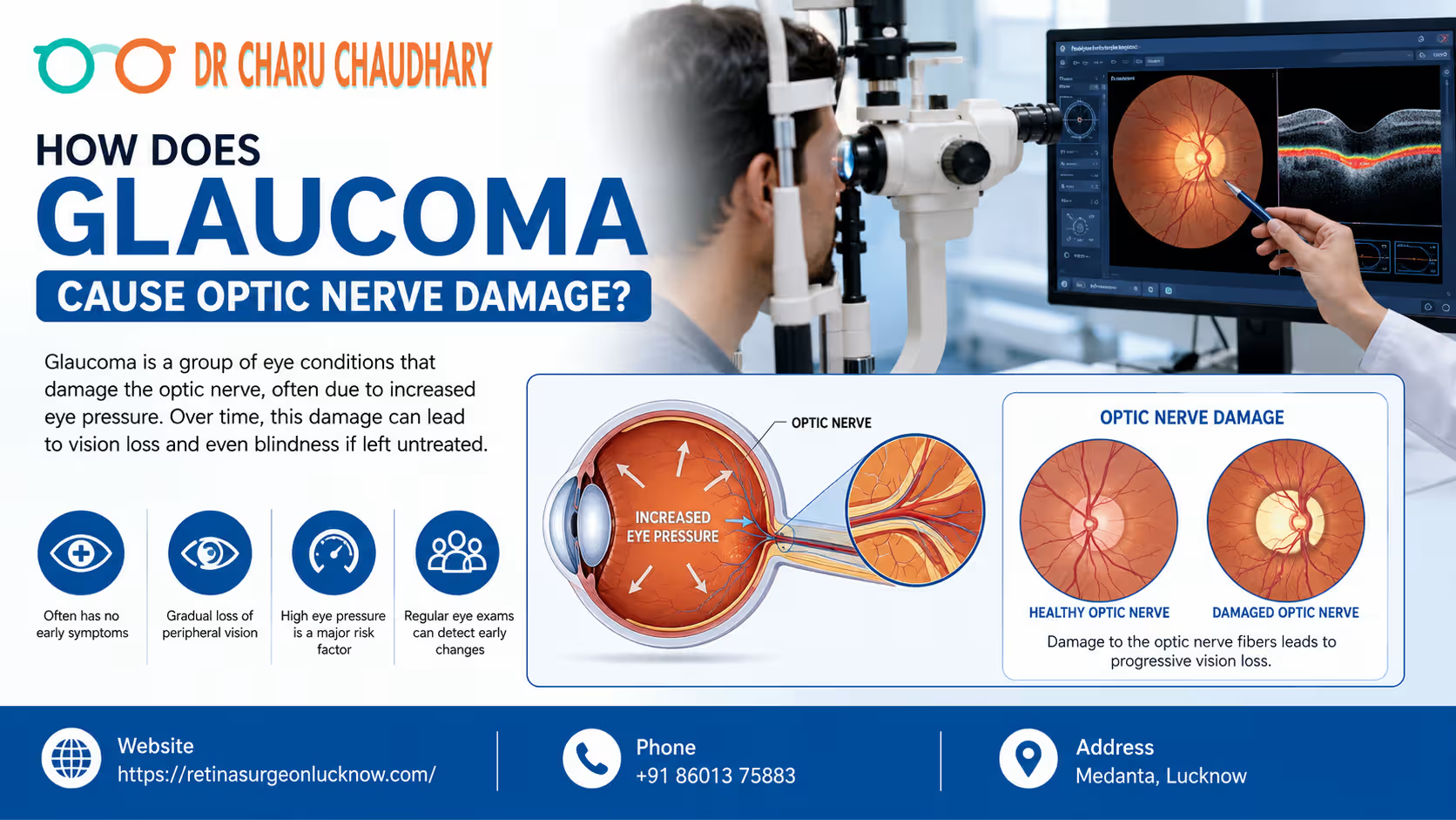
Vision is often considered our most precious sense, yet many of us take it for granted until it begins to fade. Among the various conditions that can threaten our sight, glaucoma stands out as one of the most mysterious and dangerous. Often called the “silent thief of sight,” glaucoma is a group of eye conditions that damage the optic nerve—the vital link between your eye and your brain. Because this damage often happens slowly and without pain, millions of people worldwide are unaware they even have the condition until significant vision loss has occurred. Understanding the relationship between glaucoma and optic nerve health is the first step toward prevention. In this comprehensive guide, we will explore the mechanics of glaucoma optic nerve damage, why early detection is life-changing, and how experts like Dr. Charu Chaudhary are helping patients preserve their vision. How Glaucoma Damages the Optic Nerve Glaucoma causes optic nerve damage by increasing pressure inside the eye or reducing blood supply to the nerve fibers. Over time, this pressure crushes sensitive nerve cells, leading to permanent vision loss if the condition is not detected and treated early. What Is Glaucoma? Understanding the Basics At its core, glaucoma is not just a single disease but a category of ocular disorders characterized by progressive damage to the optic nerve. Understanding Glaucoma in Simple Terms Think of your eye like a sink with a faucet and a drain. The “faucet” produces a clear fluid called aqueous humor to nourish the eye. The “drain” (located at the angle where the iris and cornea meet) allows this fluid to leave. In a healthy eye, the production and drainage are balanced. In glaucoma, the drain gets clogged or works inefficiently, causing fluid to build up. This buildup increases pressure, which eventually pushes against the optic nerve. How Common Is Glaucoma? Glaucoma is a leading cause of irreversible blindness globally. According to the World Health Organization (WHO), it is the second leading cause of blindness after cataracts. However, unlike cataracts, which can be surgically “cured” to restore sight, the vision loss caused by glaucoma is permanent. Why Glaucoma Is a Serious Eye Disease The danger of glaucoma lies in its stealthy nature. In the most common form (open-angle glaucoma), there are no symptoms in the early stages. No pain, no redness, and no sudden blurring. By the time a patient notices a “tunnel vision” effect, up to 40% of the optic nerve fibers may already be destroyed. This is why Dr. Charu Chaudhary emphasizes that regular screenings are the only way to catch the thief before it steals your sight. Can Glaucoma Cause Permanent Blindness? Yes. If left untreated, glaucoma eventually destroys the entire optic nerve, resulting in total blindness. However, with modern medical interventions and early diagnosis by the Best Eye Specialist in Lucknow, the vast majority of patients can maintain functional vision for the rest of their lives. What Is the Optic Nerve and Why Is It Important? To understand glaucoma optic nerve damage, we must first understand what the optic nerve does. How the Optic Nerve Connects the Eye to the Brain The optic nerve is often described as the “electric cable” of the eye. It is composed of more than a million tiny nerve fibers (retinal ganglion cells). These fibers collect visual information from the retina (the light-sensitive tissue at the back of the eye) and transmit it to the brain. How Visual Signals Travel Why Healthy Optic Nerves Are Essential for Vision Without a functional optic nerve, the eye and the brain cannot communicate. Even if your eye is perfectly healthy in every other way—clear lens, healthy retina, perfect cornea—you will be blind if the optic nerve is severed or destroyed. It is the “bridge” of sight. What Happens When the Optic Nerve Gets Damaged? When the fibers within the optic nerve begin to die, the “cable” loses its ability to transmit full images. Initially, the brain compensates for small gaps in the visual field. However, as more fibers die, the gaps become larger, leading to permanent blind spots. How Does Glaucoma Cause Optic Nerve Damage? The process of damage is complex and can involve several biological mechanisms. 1. Increased Eye Pressure (Intraocular Pressure – IOP) High intraocular pressure is the most significant risk factor for glaucoma. When fluid (aqueous humor) cannot drain properly, the pressure inside the eye rises. This pressure exerts physical force on the optic nerve head (the point where the nerve leaves the eye). Over time, this mechanical stress compresses the nerve fibers and the tiny blood vessels that nourish them. 2. Reduced Blood Supply to the Optic Nerve Some patients develop glaucoma even with “normal” eye pressure. This suggests that poor blood flow (ischemia) to the optic nerve also plays a role. If the blood vessels supplying the nerve are narrow or if blood pressure is too low, the nerve cells don’t get enough oxygen and nutrients, leading to cell death. 3. Damage to Retinal Nerve Fibers The optic nerve is made of the axons of retinal ganglion cells. Glaucoma specifically targets these cells. The high pressure or low blood flow triggers a process called “apoptosis” or programmed cell death. Once these cells die, they do not regenerate. 4. Progressive Loss of Nerve Cells The damage usually starts at the outer edges of the optic nerve, which corresponds to our peripheral (side) vision. As the disease progresses, the damage moves inward toward the center, eventually affecting central vision and leading to total blindness. Why the Damage Is Usually Permanent Unlike skin or bone, the nerve cells in the human central nervous system (which includes the optic nerve) do not have the capacity to regrow once they are dead. This is why glaucoma treatment focuses on “saving what’s left” rather than “restoring what’s lost.” The Step-by-Step Process of Glaucoma Damage Glaucoma is a journey of progression. Understanding these stages can help patients realize where they stand. Stage What is Happening? Impact on Vision Stage 1: Increased Pressure Fluid drainage slows down; IOP begins to rise. No noticeable

Diabetic Retinopathy Treatment in Lucknow: A Patient Guide Poasted by Dr Charu Chaudhary | Retina Specialist in Lucknow If you or a family member has been living with diabetes for a long time, you know that keeping your blood sugar in check is a daily job. But did you know that diabetes can slowly and quietly damage your eyesight? This condition is called Diabetic Retinopathy. Living in a busy city like Lucknow, we often ignore small health issues until they become big problems. However, when it comes to your eyes, waiting is not an option. If you are looking for the most reliable Diabetic Retinopathy Treatment, this guide will explain everything you need to know in simple words. We will also talk about how the best eye surgeon in Lucknow, Dr Charu Chaudhary, can help you save your vision. What Exactly is Diabetic Retinopathy? Let’s break it down into simple terms. Your eye is like a camera. The back of the eye has a “film” called the retina. This retina captures images and sends them to your brain. To work properly, the retina needs a constant supply of blood through tiny blood vessels. When your blood sugar stays high for too long, these tiny blood vessels get blocked or damaged. This whole process is what we call Diabetic Retinopathy. If left untreated, it can lead to permanent blindness. Why Should You Seek Treatment in Lucknow? Lucknow has become a major hub for medical care in North India. Earlier, people used to travel to Delhi or Mumbai for advanced eye surgeries. Today, Lucknow offers world-class technology and experienced doctors. Whether you are looking for a routine check-up or advanced Diabetic Retinopathy Treatment, the city has clinics equipped with the latest lasers and scanning machines. Choosing a local expert like Dr Charu Chaudhary means you get international-standard care without the stress of traveling to another state. Symptoms: When Should You See the Best Eye Surgeon in Lucknow? In the early stages, you might not feel any pain or notice any change in your vision. This is why it is often called a “silent thief.” However, as the damage increases, you may notice: If you notice any of these, you must immediately book a consultation with the best eye surgeon in Lucknow. Understanding the Stages: A Quick Comparison It helps to know which stage of the disease you are in. Your doctor will use this chart to explain your condition: Stage Name What is happening inside the eye? Risk Level Mild NPDR Tiny bulges in blood vessels; minor leakage. Low (Monitor) Moderate NPDR Some vessels are blocked; swelling starts. Medium Severe NPDR Many vessels are blocked; blood supply is cut off. High (Requires Treatment) Proliferative (PDR) New, fragile vessels grow and bleed into the eye. Very High (Emergency) Macular Edema The center of the retina (Macula) swells up. High (Blurry Vision) Meet Your Specialist: Dr Charu Chaudhary When it comes to something as delicate as your eyes, you cannot settle for anything but the best. Dr Charu Chaudhary is widely regarded as the best eye surgeon in Lucknow for retinal disorders. With years of specialized training and a compassionate approach, Dr. Chaudhary helps patients navigate the complexities of diabetes-related eye issues. Her clinic is known for using the latest diagnostic tools to catch the disease early, which is the most important part of successful Diabetic Retinopathy Treatment. Step-by-Step: What Happens During Your First Visit? If it is your first time visiting a specialist for Diabetic Retinopathy Treatment, you might feel nervous. Here is a step-by-step guide on what to expect: Step 1: Vision Check The staff will ask you to read letters from a chart to see how much your vision has changed. Step 2: Dilation The doctor will put special drops in your eyes. These drops make your pupils grow large. This allows the surgeon to see the back of your eye clearly. Note: Your vision will be blurry for 3–4 hours after this. Step 3: Retinal Examination Using a bright light and a special lens, Dr Charu Chaudhary will look at your retina to check for leaks, fatty deposits, or new blood vessel growth. Step 4: Digital Imaging (OCT Scan) This is like an ultrasound for the eye. It creates a 3D map of your retina to show exactly where the swelling is located. Step 5: Discussion and Planning Based on the results, the doctor will sit with you and explain the best course of action. Modern Options for Diabetic Retinopathy Treatment The good news is that medical science has advanced rapidly. We now have several ways to stop vision loss. 1. Laser Treatment (Photocoagulation) This is a very common procedure. A tiny, powerful beam of light is used to seal leaking blood vessels. It also helps shrink the abnormal new blood vessels. It is usually done in the clinic and doesn’t require an overnight stay. 2. Anti-VEGF Injections This might sound scary, but it is one of the most effective treatments today. The doctor injects a special medicine into the eye (after numbing it completely). This medicine blocks the chemicals that cause swelling and new vessel growth. Many patients see a significant improvement in vision after a few sessions. 3. Vitrectomy (Surgery) If there is a lot of blood in the center of the eye or if the retina is starting to pull away, you may need a vitrectomy. This is a surgery where the best eye surgeon in Lucknow removes the blood-filled gel from the eye and replaces it with a clear liquid or gas. Why Dr Charu Chaudhary is the Right Choice Choosing Dr Charu Chaudhary means you are choosing expertise combined with advanced technology. Here is why she stands out: How to Prepare for Your Treatment Day If you are scheduled for a procedure, follow these simple steps: Lifestyle Tips: Protecting Your Eyes at Home Treatment is only 50% of the battle. The other 50% depends on how you manage your health at home. Frequently Asked Questions (FAQs) 1. Can Diabetic Retinopathy be reversed? Damage to the nerves that has already occurred is hard to reverse. However, Diabetic Retinopathy