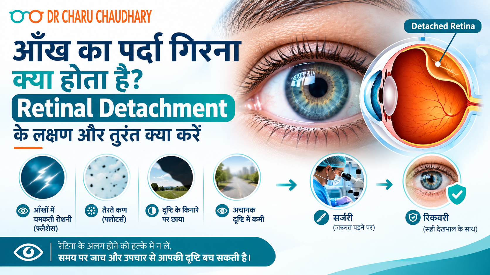
कल्पना कीजिए – आप सुबह उठते हैं और अचानक महसूस होता है कि एक आँख के सामने काला पर्दा गिर रहा है। या शायद रात में आपको बिजली जैसी चमक दिखी और सोचा “थकान होगी।” अगले दिन उसी आँख से धुंधला दिखने लगा। अधिकतर लोग इसे नज़रअंदाज़ कर देते हैं। यही सबसे बड़ी गलती है। जो आप महसूस कर रहे थे, वह Retinal Detachment – यानी आँख का पर्दा गिरना – के शुरुआती संकेत थे। और यह एक ऐसी medical emergency है जिसमें हर घंटा महत्वपूर्ण होता है। मैं Dr. Charu Chaudhary हूँ, Lucknow के Medanta Hospital में Vitreoretinal Surgeon। अपने वर्षों के अनुभव में मैंने देखा है कि जो मरीज़ 24–48 घंटे के भीतर आते हैं, उनकी रोशनी बचाई जा सकती है। जो देर करते हैं – उनके लिए रास्ता बहुत मुश्किल हो जाता है। इस blog में मैं आपको सरल भाषा में बताऊँगी कि आँख का पर्दा गिरना क्या होता है, इसके लक्षण क्या हैं, और सबसे ज़रूरी – इस स्थिति में आपको तुरंत क्या करना चाहिए। रेटिना क्या होती है और वह इतनी ज़रूरी क्यों है? आँख को एक camera की तरह समझिए। जिस तरह camera में film होती है जो तस्वीर कैद करती है – उसी तरह आँख में retina होती है। यह एक पतली, नाज़ुक परत होती है जो आँख के पिछले हिस्से में होती है। जब भी कोई रोशनी आँख में आती है, रेटिना उसे electrical signals में बदलता है और इन signals को Optic Nerve के ज़रिए दिमाग तक पहुँचाता है। दिमाग इन्हें image के रूप में पहचानता है – और आपको दिखता है। अगर यही रेटिना अपनी जगह से हट जाए – तो signal टूट जाता है। दृष्टि धुंधली हो जाती है, गायब हो सकती है, और बिना इलाज के हमेशा के लिए जा सकती है। आँख का पर्दा गिरता क्यों है – मुख्य कारण Retinal Detachment अचानक होता है, लेकिन इसके पीछे कुछ जोखिम कारण होते हैं जिनके बारे में आपको पहले से जानकारी होनी चाहिए: चोट या Trauma: आँख पर तेज़ चोट लगने से, पटाखे फूटने से, या खेल के दौरान चोट लगने से रेटिना खिंच सकती है और अपनी जगह से हट सकती है। High Myopia (तेज़ चश्मा): जिन लोगों का नंबर -6 या उससे अधिक है, उनकी आँख का आकार सामान्य से बड़ा होता है। इससे रेटिना पर खिंचाव रहता है और detachment का खतरा बढ़ जाता है। Diabetes: लम्बे समय से बेकाबू शुगर रेटिना की नसों को कमज़ोर करती है। इसे Diabetic Retinopathy कहते हैं – और यह Retinal Detachment का एक बड़ा कारण बन सकती है। बढ़ती उम्र: उम्र के साथ आँख के अंदर का gel (Vitreous) सिकुड़ता है और रेटिना को खींच सकता है। 50 वर्ष से अधिक उम्र के लोगों में यह ज़्यादा देखा जाता है। पहले का Retinal Detachment: एक आँख में हो चुका है तो दूसरी आँख में भी जोखिम बढ़ जाता है। पारिवारिक इतिहास: परिवार में किसी को हो चुका है तो आपको नियमित जाँच करानी चाहिए। आँख का पर्दा गिरने के लक्षण – जिन्हें कभी नज़रअंदाज़ न करें यह वह हिस्सा है जिसे मैं सबसे ज़्यादा ज़ोर देकर बताना चाहती हूँ। Retinal Detachment में दर्द नहीं होता—यही इसे इतना खतरनाक बनाता है। लोग सोचते हैं “दर्द नहीं तो गंभीर नहीं होगा।” यह सोच गलत है। इन लक्षणों में से कोई भी दिखे – तुरंत doctor के पास जाएँ: अचानक बहुत सारे काले धब्बे या जाले दिखना (Floaters) हाँ, कुछ floaters सामान्य हो सकते हैं। लेकिन अगर अचानक बहुत सारे नए floaters एक साथ दिखें – जैसे मकड़ी का जाला या काली बिंदियाँ तैर रही हों – तो यह रेटिना में खिंचाव या टूटन का संकेत है। बिजली जैसी चमक दिखना (Flashes of Light) आँखें बंद करने पर या अंधेरे में भी चमक दिखे – जैसे camera flash हो। यह इस बात का संकेत है कि Vitreous रेटिना को खींच रहा है। आँख के सामने काला पर्दा या छाया आना ऐसा महसूस हो जैसे किसी ने एक तरफ से curtain खींच दिया हो। पहले side vision जाती है, फिर धीरे-धीरे बीच का हिस्सा भी ढकने लगता है। यह सबसे serious लक्षण है। इस स्थिति में हर मिनट मायने रखता है। अचानक धुंधला दिखना, सिर्फ एक आँख से अचानक एक आँख से कम या धुंधला दिखना – और यह चश्मा लगाने पर भी ठीक न हो। यह इतनी बड़ी Emergency क्यों है? रेटिना एक जीवित tissue है। जब यह अपनी जगह से हट जाती है, तो उसे blood supply मिलनी बंद हो जाती है। जिस तरह खून न मिलने पर कोई भी अंग मरने लगता है – उसी तरह रेटिना की कोशिकाएँ भी मरने लगती हैं। पहले 24 घंटे: अगर पर्दा centre (Macula) तक नहीं पहुँचा, तो surgery के बाद अच्छी रोशनी वापस आने की संभावना बहुत अधिक है। 24–72 घंटे: Macula तक पहुँचने के बाद रोशनी वापस आना मुश्किल होने लगता है। 72 घंटे से अधिक: रेटिना की कोशिकाएँ स्थायी रूप से मर सकती हैं। रोशनी का नुकसान permanent हो सकता है। इसीलिए Retinal Detachment को हम “Golden Hour Emergency” कहते हैं। तुरंत क्या करें – Step by Step अगर आपको या आपके किसी परिवार वाले को ऊपर दिए गए लक्षण दिखें: Step 1: आँख को मलें नहीं, दबाएँ नहीं। Step 2: दोनों आँखें बंद करके शांत रहें – ज़्यादा eye movement से रेटिना खिंच सकता है। Step 3: कोई भी घरेलू उपाय, eye drop, या “देखते हैं कल” – यह सब बंद करें। Step 4: तुरंत किसी Vitreoretinal Specialist से मिलें। General physician या chemist के पास जाने का वक्त नहीं है। Lucknow में हैं तो अभी appointment book करें – हम emergency cases को उसी दिन देखते हैं। Diagnosis कैसे होती है? जब आप clinic आते हैं, तो हम कुछ ज़रूरी जाँचें करते हैं: Dilated Fundus Examination: आँख की पुतली को drops से बड़ा करके रेटिना को सीधे देखा जाता है। B-Scan Ultrasound: अगर आँख के अंदर खून है या साफ नहीं दिख रहा, तो Ultrasound से रेटिना की स्थिति देखी जाती है। OCT (Optical Coherence Tomography): रेटिना की परतों की detailed imaging – यह बताता है कि कितना नुकसान हुआ है। ये सब जाँचें दर्द रहित हैं
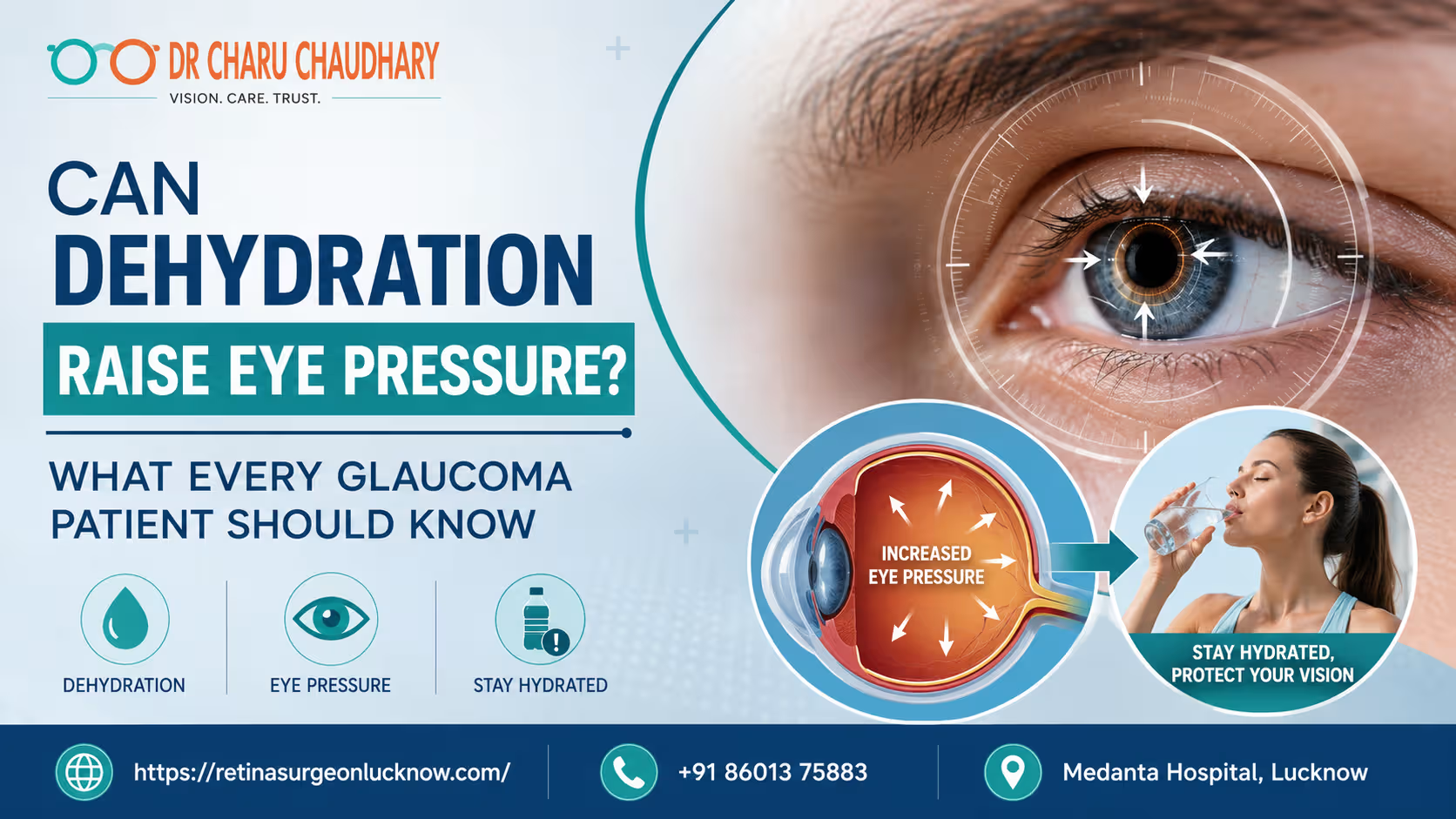
Dehydration may temporarily influence intraocular pressure in some people and can significantly affect overall eye health. While dehydration alone does not usually cause glaucoma, maintaining proper hydration is important for patients already diagnosed with the condition. Fluctuations in body fluids can impact how the eye regulates its internal pressure and supports the optic nerve. Can Not Drinking Enough Water Affect Your Eye Pressure? Maintaining the right fluid balance is vital for your health. Most people know that dehydration affects the heart and kidneys. However, many patients are surprised to learn that it also affects their eyes. For individuals living with glaucoma, understanding the link between hydration and eye pressure is essential. Eye pressure, or intraocular pressure (IOP), is the primary metric used to manage glaucoma. When your body lacks sufficient water, your systemic circulation changes. These changes can impact the delicate structures inside your eye. Consequently, your vision may face unnecessary risks if you ignore your daily water intake. Dr. Charu Chaudhary, an expert in ocular health, emphasizes that hydration is a pillar of preventative care. She often advises that stable fluid levels help maintain a stable environment for the optic nerve. If you are looking for the Best Eye Specialist in Lucknow, finding a professional who looks at your systemic health is key. Why Glaucoma Patients Should Pay Attention to Hydration Glaucoma is often called the “silent thief of sight.” It slowly damages the optic nerve, usually due to high internal eye pressure. Because this condition is so sensitive to pressure changes, even minor fluctuations matter. Proper hydration ensures that the blood flowing to your eyes is rich in oxygen and nutrients. When you are dehydrated, your blood can become more viscous. This change may reduce the efficiency of blood flow to the optic nerve. Therefore, staying hydrated isn’t just about the “pressure number” on a chart. It is about protecting the nerve fibers that allow you to see. Is there a real connection between dehydration and eye health? Medical research suggests that while severe dehydration might actually lower eye pressure slightly, the secondary effects are harmful. Dry eyes, blurred vision, and poor circulation can worsen the overall experience of living with glaucoma. What Is Eye Pressure (Intraocular Pressure)? Intraocular pressure is the fluid pressure inside your eye. Your eye produces a clear fluid called aqueous humor. This fluid nourishes the front part of the eye and maintains its shape. To keep pressure steady, the eye must drain as much fluid as it produces. When the drainage system becomes clogged or inefficient, the fluid builds up. This buildup increases the pressure within the eyeball. Think of it like a balloon being overfilled with water. If the pressure remains too high for too long, it pushes against the back of the eye. How Is Eye Pressure Measured? Eye specialists use a process called tonometry to measure IOP. During an exam, the doctor will use a device to gently touch or blow a puff of air onto the surface of your eye. This measurement is recorded in millimeters of mercury (mmHg). A normal eye pressure range is generally between 12 and 21 mmHg. However, “normal” can vary from person to person. Some people develop glaucoma with low pressure, while others have high pressure without nerve damage. This is why regular monitoring is so crucial for long-term vision. Why Is Eye Pressure Important for Vision? Your optic nerve is the “cable” that connects your eye to your brain. It is made of over a million tiny nerve fibers. High eye pressure compresses these fibers. Over time, the fibers begin to die off, leading to permanent vision loss. Because these fibers do not regenerate, the damage is irreversible. Managing eye pressure is the only proven way to stop or slow the progression of glaucoma. Consistency in your habits, including your water intake, helps keep these pressure levels within a safe range. What Is Glaucoma and How Does It Damage Vision? Glaucoma is a group of eye conditions that lead to optic nerve damage. It is typically categorized by how the fluid drainage system is blocked. The most common form is Open-Angle Glaucoma, where the drainage angle looks open but functions poorly. Another form is Angle-Closure Glaucoma, which can occur suddenly and constitutes a medical emergency. Regardless of the type, the result is often the same: peripheral vision fades first. If left untreated, the damage moves inward, eventually leading to total blindness. High eye pressure is the most significant modifiable risk factor for this disease. By controlling this pressure through medication, surgery, and lifestyle choices, patients can preserve their sight for a lifetime. This is why consulting the Best Eye Surgeon in Lucknow is a vital step for those at risk. Can Dehydration Really Raise Eye Pressure? The relationship between dehydration and eye pressure is complex. In many cases, severe systemic dehydration actually leads to a decrease in intraocular pressure. This happens because the body produces less aqueous humor when it is trying to conserve water. However, the problem arises when the body is in a state of flux. Chronic dehydration can lead to “dry eye” symptoms that mimic or worsen glaucoma discomfort. Furthermore, when a dehydrated person suddenly drinks a massive amount of water, it can cause a temporary spike in eye pressure. What Does Medical Research Say? Research indicates that the body’s “osmolality”—the concentration of particles in your blood—affects the fluid in your eyes. When you are dehydrated, your blood is more concentrated. This can pull fluid out of the eye, lowering pressure. Conversely, “water loading” (drinking a liter of water in under five minutes) is a known test to see if eye pressure will spike. For a glaucoma patient, these rapid shifts are dangerous. Stability is the goal. Therefore, avoiding extreme dehydration is just as important as avoiding rapid over-hydration. Is the Effect Temporary or Long-Term? Most pressure changes related to hydration are temporary. However, the cumulative effect of poor hydration on the optic nerve’s blood supply can be long-term. Your eyes need consistent hydration
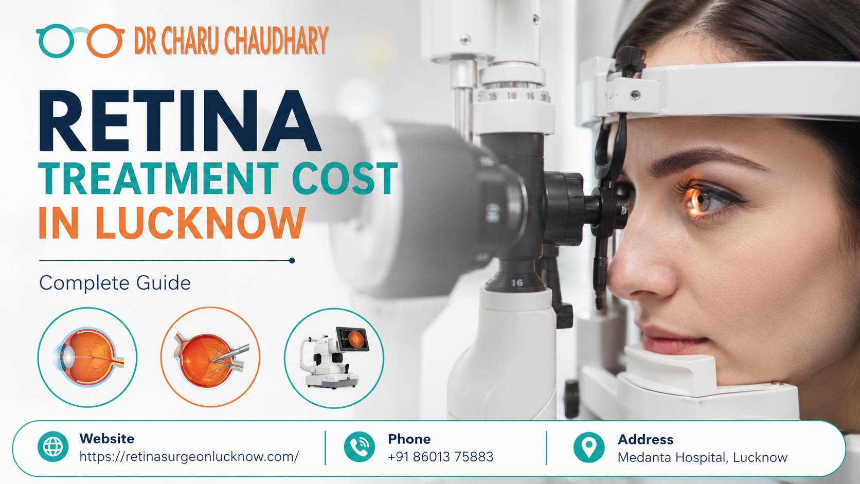
In recent years, the prevalence of retinal diseases has seen a significant surge across India, with Lucknow being no exception. As a major healthcare hub in Uttar Pradesh, Lucknow witnesses thousands of patients daily seeking specialized care for vision-threatening conditions. The retina, a thin layer of light-sensitive tissue at the back of the eye, acts much like the film in a traditional camera. When it becomes damaged due to age, diabetes, or trauma, the consequences can be devastating, often leading to irreversible vision loss if left untreated. One of the primary concerns for patients and their families when facing a retinal diagnosis is the financial aspect. “How much will the treatment cost?” is usually the first question asked after a diagnosis is confirmed. Understanding the Retina Treatment Cost is essential for making informed decisions, planning finances, and ensuring that quality care is not delayed. Early intervention is not just a clinical necessity; it is a financial strategy. Detecting a retinal tear early might cost a few thousand rupees for a laser procedure, whereas ignoring it could lead to a retinal detachment requiring surgery costing tens of thousands. Retina Treatment Cost in Lucknow varies depending on the retinal condition, diagnostic tests, treatment method, and whether surgery is required. Early diagnosis often reduces complications, treatment complexity, and overall healthcare expenses, ensuring better visual outcomes at a lower cost. Quick Facts: Retina Treatment at a Glance Service/Treatment Approximate Cost Range (INR) Recovery Timeline Consultation (Retina Specialist) ₹500 – ₹1,500 Immediate OCT Scan (Optical Coherence Tomography) ₹1,500 – ₹3,500 Immediate Fundus Photography/Angiography ₹2,000 – ₹5,000 1 – 2 Hours Retina Laser Treatment (per session) ₹5,000 – ₹15,000 1 – 2 Days Anti-VEGF Injection (per dose) ₹7,000 – ₹60,000 1 – 3 Days Retinal Detachment Surgery ₹40,000 – ₹1,20,000 2 – 6 Weeks Vitrectomy Surgery ₹50,000 – ₹1,50,000 3 – 8 Weeks What Is the Retina and Why Is It Important? Understanding the Role of the Retina The retina is arguably the most vital part of the eye’s anatomy. It is a complex, multi-layered structure that captures light entering the eye and converts it into electrical signals. These signals are then transmitted via the optic nerve to the brain, which interprets them as images. Without a functioning retina, the eye may be structurally intact, but the “vision” process cannot occur. How the Retina Helps You See Think of the retina as the “sensor” of your eye. The central part of the retina, known as the macula, is responsible for sharp, detailed, central vision. This allows you to read, drive, and recognize faces. The peripheral retina provides you with side vision, helping you navigate your surroundings. What Happens When the Retina Is Damaged? Unlike some other tissues in the body, the retina has a very limited capacity to regenerate. Damage caused by high blood sugar (diabetes), high blood pressure, or physical trauma can lead to scarring, bleeding, or detachment. When the retina is damaged, images become blurred, distorted, or vanish entirely. Because the retina is directly connected to the brain’s visual cortex, any delay in treatment can lead to permanent atrophy of the nerve fibers. Common Retina Conditions That Require Treatment Understanding the specific condition you have is the first step in estimating the retinal disease treatment cost. Different pathologies require vastly different approaches. 1. Diabetic Retinopathy This is the leading cause of blindness among working-age adults. Chronic high blood sugar damages the tiny blood vessels inside the retina. 2. Retinal Detachment A medical emergency where the retina pulls away from its underlying layer of support tissue. 3. Age-Related Macular Degeneration (AMD) Common in individuals over 50, AMD affects the macula. 4. Retinal Vein Occlusion (RVO) A blockage of the small veins that carry blood away from the retina. This often causes sudden, painless vision loss in one eye and requires injections or laser treatment to manage swelling (edema). 5. Macular Edema This refers to the swelling of the macula, often a complication of diabetes or RVO. The eye retina treatment for this usually involves a series of injections. 6. Retinal Tears and Holes Small breaks in the retina can lead to detachment. These are often treated quickly with “green laser” therapy to seal the edges of the tear. Retina Treatment Cost in Lucknow: Overview Lucknow offers a wide spectrum of eye care, ranging from government-run charitable hospitals to ultra-modern private eye centers. The Retina treatment price in Lucknow is generally more affordable than in Delhi or Mumbai, but the quality of technology and surgical expertise remains world-class. Average Cost Range A patient can expect to spend anywhere from ₹5,000 for a minor laser procedure to ₹1,50,000 for a complex vitreoretinal surgery involving imported silicon oil or gases. Why Treatment Costs Vary It is difficult to provide a single “price tag” for retina care because no two eyes are the same. A Retina specialist in Lucknow will determine the price based on: Diagnostic Investigation Costs Accurate diagnosis is the foundation of successful treatment. Before any procedure, a Retina Specialist in Lucknow will perform several tests to map the extent of the damage. Diagnostic Test Purpose Estimated Cost (INR) Consultation Fee Expert evaluation of the eye ₹500 – ₹1,500 OCT Scan Cross-sectional imaging of the retina ₹1,500 – ₹3,500 Fundus Fluorescein Angiography (FFA) Mapping blood flow with dye ₹2,500 – ₹4,500 B-Scan Ultrasound Seeing the retina when cataracts are present ₹800 – ₹1,500 Indocyanine Green Angiography (ICG) Visualizing deeper choroidal vessels ₹5,000 – ₹8,000 Optical Coherence Tomography Angiography (OCTA) Non-invasive blood flow mapping ₹3,000 – ₹6,000 The retina checkup cost usually includes a dilated eye exam and at least one imaging test, like an OCT. Factors That Affect Retina Treatment Cost When budgeting for your care, consider these influencing factors: Anti-VEGF Injection Cost in Lucknow Anti-VEGF (Vascular Endothelial Growth Factor) injections have revolutionized retina care. They “dry up” leaking vessels and are the gold standard for Wet AMD and Diabetic Macular Edema. What Are Anti-VEGF Injections? These are medications injected directly into the vitreous (the gel-like substance in the eye). The procedure is done under local anesthesia and takes less than
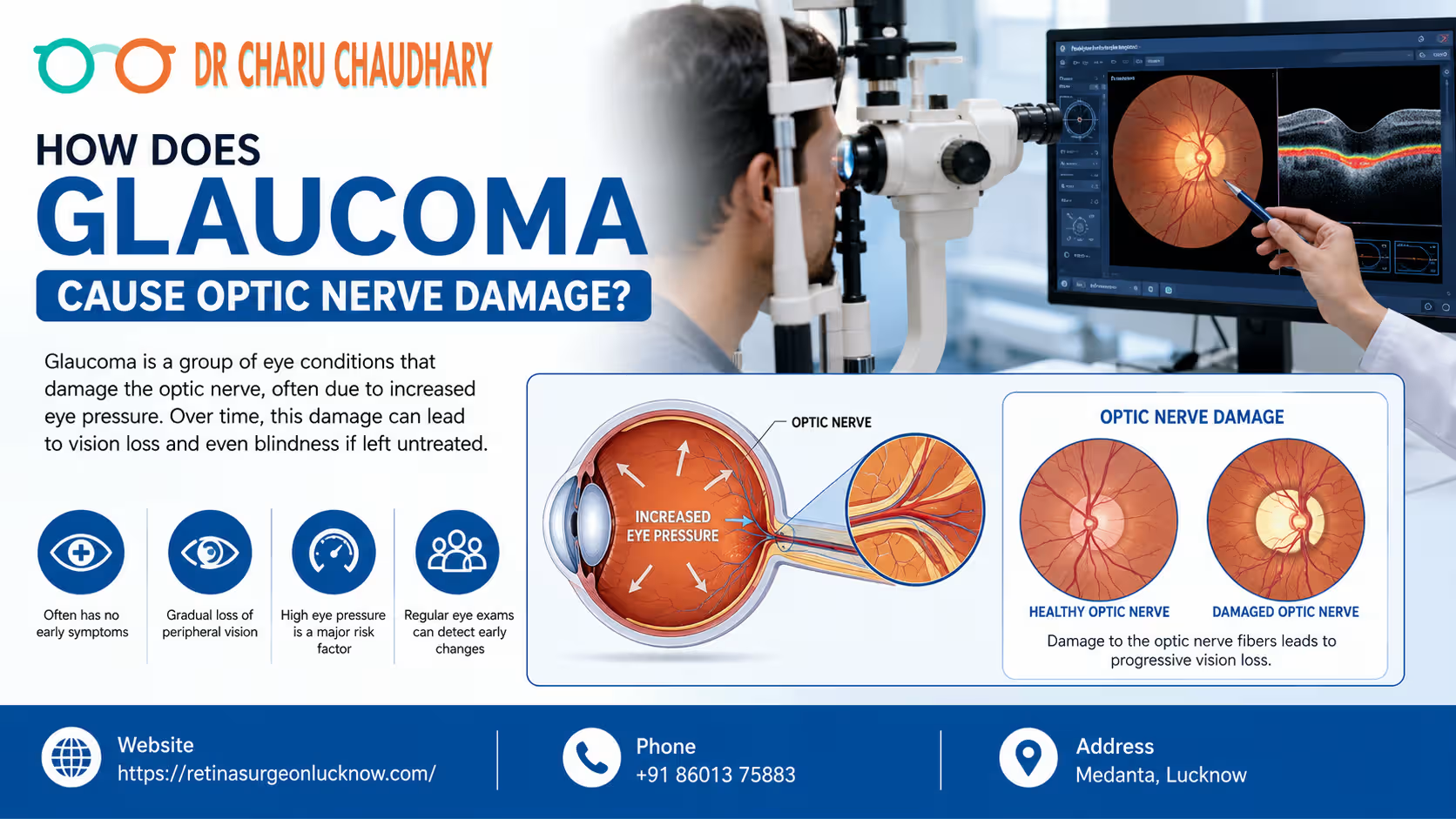
Vision is often considered our most precious sense, yet many of us take it for granted until it begins to fade. Among the various conditions that can threaten our sight, glaucoma stands out as one of the most mysterious and dangerous. Often called the “silent thief of sight,” glaucoma is a group of eye conditions that damage the optic nerve—the vital link between your eye and your brain. Because this damage often happens slowly and without pain, millions of people worldwide are unaware they even have the condition until significant vision loss has occurred. Understanding the relationship between glaucoma and optic nerve health is the first step toward prevention. In this comprehensive guide, we will explore the mechanics of glaucoma optic nerve damage, why early detection is life-changing, and how experts like Dr. Charu Chaudhary are helping patients preserve their vision. How Glaucoma Damages the Optic Nerve Glaucoma causes optic nerve damage by increasing pressure inside the eye or reducing blood supply to the nerve fibers. Over time, this pressure crushes sensitive nerve cells, leading to permanent vision loss if the condition is not detected and treated early. What Is Glaucoma? Understanding the Basics At its core, glaucoma is not just a single disease but a category of ocular disorders characterized by progressive damage to the optic nerve. Understanding Glaucoma in Simple Terms Think of your eye like a sink with a faucet and a drain. The “faucet” produces a clear fluid called aqueous humor to nourish the eye. The “drain” (located at the angle where the iris and cornea meet) allows this fluid to leave. In a healthy eye, the production and drainage are balanced. In glaucoma, the drain gets clogged or works inefficiently, causing fluid to build up. This buildup increases pressure, which eventually pushes against the optic nerve. How Common Is Glaucoma? Glaucoma is a leading cause of irreversible blindness globally. According to the World Health Organization (WHO), it is the second leading cause of blindness after cataracts. However, unlike cataracts, which can be surgically “cured” to restore sight, the vision loss caused by glaucoma is permanent. Why Glaucoma Is a Serious Eye Disease The danger of glaucoma lies in its stealthy nature. In the most common form (open-angle glaucoma), there are no symptoms in the early stages. No pain, no redness, and no sudden blurring. By the time a patient notices a “tunnel vision” effect, up to 40% of the optic nerve fibers may already be destroyed. This is why Dr. Charu Chaudhary emphasizes that regular screenings are the only way to catch the thief before it steals your sight. Can Glaucoma Cause Permanent Blindness? Yes. If left untreated, glaucoma eventually destroys the entire optic nerve, resulting in total blindness. However, with modern medical interventions and early diagnosis by the Best Eye Specialist in Lucknow, the vast majority of patients can maintain functional vision for the rest of their lives. What Is the Optic Nerve and Why Is It Important? To understand glaucoma optic nerve damage, we must first understand what the optic nerve does. How the Optic Nerve Connects the Eye to the Brain The optic nerve is often described as the “electric cable” of the eye. It is composed of more than a million tiny nerve fibers (retinal ganglion cells). These fibers collect visual information from the retina (the light-sensitive tissue at the back of the eye) and transmit it to the brain. How Visual Signals Travel Why Healthy Optic Nerves Are Essential for Vision Without a functional optic nerve, the eye and the brain cannot communicate. Even if your eye is perfectly healthy in every other way—clear lens, healthy retina, perfect cornea—you will be blind if the optic nerve is severed or destroyed. It is the “bridge” of sight. What Happens When the Optic Nerve Gets Damaged? When the fibers within the optic nerve begin to die, the “cable” loses its ability to transmit full images. Initially, the brain compensates for small gaps in the visual field. However, as more fibers die, the gaps become larger, leading to permanent blind spots. How Does Glaucoma Cause Optic Nerve Damage? The process of damage is complex and can involve several biological mechanisms. 1. Increased Eye Pressure (Intraocular Pressure – IOP) High intraocular pressure is the most significant risk factor for glaucoma. When fluid (aqueous humor) cannot drain properly, the pressure inside the eye rises. This pressure exerts physical force on the optic nerve head (the point where the nerve leaves the eye). Over time, this mechanical stress compresses the nerve fibers and the tiny blood vessels that nourish them. 2. Reduced Blood Supply to the Optic Nerve Some patients develop glaucoma even with “normal” eye pressure. This suggests that poor blood flow (ischemia) to the optic nerve also plays a role. If the blood vessels supplying the nerve are narrow or if blood pressure is too low, the nerve cells don’t get enough oxygen and nutrients, leading to cell death. 3. Damage to Retinal Nerve Fibers The optic nerve is made of the axons of retinal ganglion cells. Glaucoma specifically targets these cells. The high pressure or low blood flow triggers a process called “apoptosis” or programmed cell death. Once these cells die, they do not regenerate. 4. Progressive Loss of Nerve Cells The damage usually starts at the outer edges of the optic nerve, which corresponds to our peripheral (side) vision. As the disease progresses, the damage moves inward toward the center, eventually affecting central vision and leading to total blindness. Why the Damage Is Usually Permanent Unlike skin or bone, the nerve cells in the human central nervous system (which includes the optic nerve) do not have the capacity to regrow once they are dead. This is why glaucoma treatment focuses on “saving what’s left” rather than “restoring what’s lost.” The Step-by-Step Process of Glaucoma Damage Glaucoma is a journey of progression. Understanding these stages can help patients realize where they stand. Stage What is Happening? Impact on Vision Stage 1: Increased Pressure Fluid drainage slows down; IOP begins to rise. No noticeable
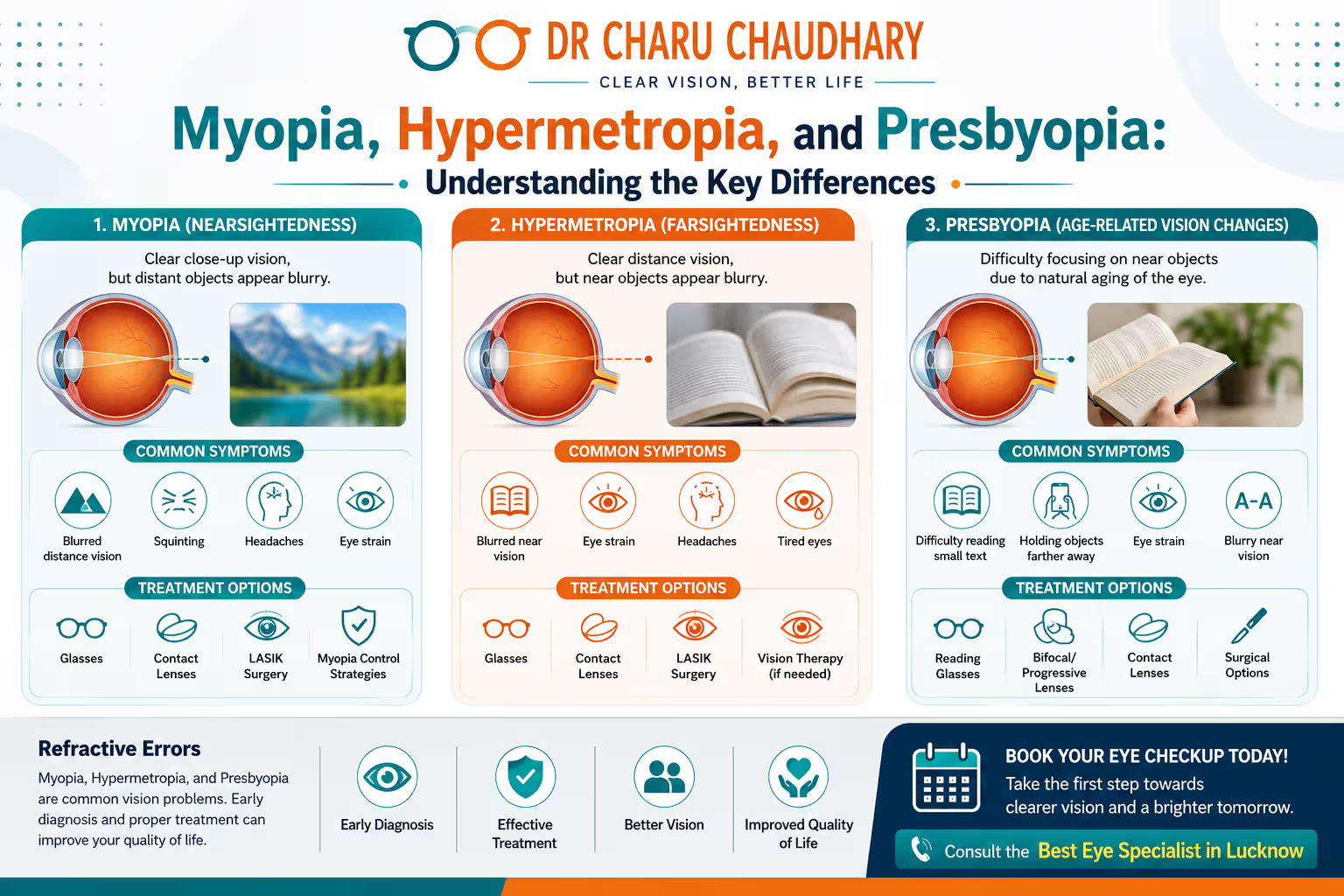
Clear vision is something many of us take for granted until things start to look a little blurry. Whether you are struggling to read the fine print on a menu, finding it hard to see road signs while driving, or noticing that your child is squinting at the television, vision changes can be unsettling. The most common reasons for blurred vision are refractive errors. While the terms Myopia, Hypermetropia, and Presbyopia might sound like complex medical jargon, they represent the three most frequent ways our eyes struggle to focus light. Understanding these conditions is the first step toward reclaiming clear sight and maintaining long-term eye health. Myopia, Hypermetropia, and Presbyopia are common refractive errors affecting how the eye focuses light. Myopia (nearsightedness) causes difficulty seeing distant objects. Hypermetropia (farsightedness) affects near vision clarity. Presbyopia is an age-related condition where the eye’s lens loses flexibility, making it difficult to focus on close-up tasks like reading. Introduction Vision is perhaps our most dominant sense, guiding how we interact with the world, learn, and work. However, according to the World Health Organization, refractive errors are the leading cause of vision impairment globally. Despite their prevalence, many people confuse these conditions, leading to delayed treatment or incorrect assumptions about their eye health. The modern lifestyle—characterized by increased screen time and less outdoor activity—has led to a surge in cases of Myopia, especially among children. On the other hand, as the global population ages, Presbyopia has become a universal experience for those over 40. Meanwhile, Hypermetropia often goes undiagnosed in children because the eye’s internal muscles work overtime to compensate, often leading to hidden eye strain. Early diagnosis is vital. Uncorrected refractive errors don’t just cause blurriness; they can lead to chronic headaches, reduced productivity, and, in children, developmental delays or “lazy eye” (amblyopia). This guide will break down the mechanics of the eye, explain the differences between these three conditions, and help you understand the path to perfect vision. How Normal Vision Works To understand what goes wrong in refractive errors, we must first understand how a “normal” eye (emmetropia) functions. Anatomy of the Eye Think of your eye as a high-tech camera. It has a protective outer layer, a lens for focusing, and a “film” or sensor at the back that captures the image. The main components involved in vision are the cornea, the lens, and the retina. Role of the Cornea and Lens Light enters the eye through the cornea, the clear, dome-shaped front surface. The cornea provides most of the eye’s optical power by bending (refracting) incoming light. Behind the cornea sits the crystalline lens, which is flexible. This flexibility allows the lens to change shape to fine-tune your focus, a process called accommodation. How Light Focuses on the Retina In a perfectly shaped eye, the cornea and lens work in harmony to bend light rays so they land precisely on a single focal point on the retina. The retina is a light-sensitive layer of tissue at the back of the eye. It converts light into neural signals and sends them via the optic nerve to the brain, which interprets them as images. What Happens When Vision Becomes Blurry Vision becomes blurry when the light does not land exactly on the retina. If the eye is too long, too short, or the cornea is too curved, the light focus lands in front of or behind the retina. This mismatch is what we call a refractive error. What Are Refractive Errors? Definition of Refractive Errors A refractive error is a type of vision problem that makes it hard to see clearly. It happens when the shape of your eye keeps light from focusing correctly on your retina. It is not a “disease” in the traditional sense, but rather an anatomical mismatch in the eye’s optical system. Why Refractive Errors Occur Refractive errors typically occur due to one of three factors: Common Types of Refractive Errors The four main types are: Impact on Daily Life Uncorrected refractive errors can make it difficult to perform everyday tasks. For a student, it means not being able to see the whiteboard. For a professional, it means digital eye strain and blurred text. For an older adult, it can mean a loss of independence when reading labels or using a phone. What Is Myopia (Nearsightedness)? Understanding Myopia Myopia, commonly known as nearsightedness, is a condition where close-up objects appear clear, but distant objects—like street signs or a movie screen—look blurry. It is the most common refractive error worldwide and is reaching epidemic levels in urban populations. Causes of Myopia Myopia occurs when the eyeball is too long relative to the focusing power of the cornea and lens. This causes light rays to focus at a point in front of the retina instead of directly on its surface. It can also be caused by a cornea that is too steeply curved. Symptoms of Myopia Risk Factors How Myopia Progresses Myopia usually starts in childhood and can progress until the early 20s as the eyeball continues to grow. High myopia (severe nearsightedness) increases the risk of serious eye conditions later in life, such as retinal detachment, cataracts, and glaucoma. Treatment Options What Is Hypermetropia (Farsightedness)? Understanding Hypermetropia Hypermetropia, or farsightedness, is a condition where distant objects are usually seen clearly, but close-up objects appear significantly blurred. However, the experience of hypermetropia varies by age; young people with mild hypermetropia may see clearly at all distances because their eyes can compensate, while older individuals may struggle with both near and far vision. Causes of Hypermetropia This occurs when the eyeball is too short or the cornea has too little curvature. As a result, light focuses at a point behind the retina. Symptoms of Hypermetropia Risk Factors Hypermetropia is often present at birth, but children frequently “outgrow” it as the eyeball lengthens during normal growth. It is highly hereditary. Complications if Left Untreated In children, significant uncorrected hypermetropia can lead to strabismus (crossed eyes) or amblyopia (lazy eye) because the brain begins to ignore the signals from the eye that is more out of focus. Treatment Options What Is
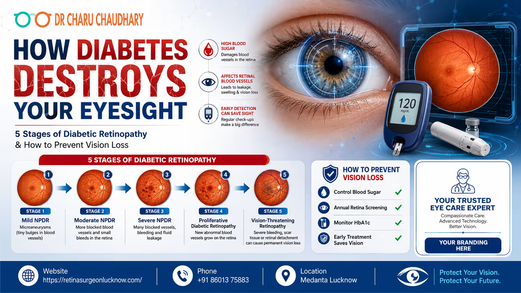
India is currently facing a dual epidemic: the explosion of diabetes and the subsequent rise in vision impairment. As the “Diabetes Capital of the World,” the burden of managing complications like diabetic retinopathy has never been more critical. This condition, often referred to as the “silent thief of sight,” remains the leading cause of preventable blindness in adults worldwide. Diabetic Retinopathy is a diabetes-related eye disease caused by damage to retinal blood vessels. Early diagnosis, good blood sugar control, and regular retina screening can help prevent severe vision loss and blindness. The Hidden Link Between Blood Sugar and Blindness The numbers are startling. According to recent health surveys, nearly one in every three people with diabetes will develop some form of eye damage. In the bustling landscape of Uttar Pradesh, particularly in cities like Lucknow, the prevalence of Type 2 diabetes is shifting toward younger age groups. This shift means people are living with high blood sugar for longer periods, significantly increasing the window for “Diabetic Vision Loss” to occur. Most patients believe that if they can see clearly, their eyes are healthy. This is a dangerous misconception. Diabetic retinopathy often begins without a single symptom. By the time vision becomes blurred or “floaters” appear, the disease has likely progressed to a stage where advanced medical intervention is required. This guide aims to bridge the gap between awareness and action, helping you understand how diabetes affects your eyes and what you can do to stop it. Key Facts About Diabetic Retinopathy To understand the gravity of this condition, let’s look at the data provided by global health leaders like the WHO, International Diabetes Federation (IDF), and the American Academy of Ophthalmology (AAO): What Is Diabetic Retinopathy? At its core, Diabetic Retinopathy is a microvascular complication. The retina is the thin layer of light-sensitive tissue at the back of your eye. It functions like the sensor in a digital camera, capturing light and converting it into electrical signals that the brain interprets as images. For the retina to function, it requires a constant and healthy supply of oxygen and nutrients through a network of tiny, delicate blood vessels. When blood sugar levels remain high for extended periods, it damages the structural integrity of these vessels. They become weak, leak fluid, or close off entirely. This process is the foundation of “Diabetic Eye Disease.” How Diabetes Affects Your Eyesight The destruction of eyesight via diabetes is a progressive, biological cascade: Why Diabetic Eye Damage Often Goes Unnoticed The human brain is remarkably good at compensating for small gaps in vision. In the early stages of retinopathy, the damage usually occurs in the peripheral (side) retina. Because your central vision remains sharp, you may not notice anything is wrong. Furthermore, diabetic eye damage does not cause pain. Unlike a “red eye” or an infection, there is no physical discomfort to alert the patient. This “silent progression” is why annual diabetic eye screening is non-negotiable for every diabetic patient, regardless of their current visual clarity. The 5 Stages of Diabetic Retinopathy Medical professionals categorize the progression of this disease to determine the appropriate treatment path. Stage 1 – Mild Non-Proliferative Diabetic Retinopathy (NPDR) This is the “alert” stage. At this point, tiny, balloon-like swellings called microaneurysms appear in the retinal blood vessels. Stage 2 – Moderate Non-Proliferative Diabetic Retinopathy As the disease advances, more blood vessels swell and lose their ability to transport blood. They may begin to leak blood and fluid, causing the retina to look “spotted” during an exam. Stage 3 – Severe Non-Proliferative Diabetic Retinopathy In this critical stage, a large number of blood vessels are blocked, depriving several areas of the retina of blood flow. These areas secrete growth factors that signal the eye to start growing new vessels. Stage 4 – Proliferative Diabetic Retinopathy (PDR) This is the advanced, vision-threatening stage. The “proliferative” part refers to the rapid growth of new, fragile blood vessels (neovascularization) along the inside surface of the retina and into the vitreous gel. Stage 5 – Advanced Vision-Threatening Diabetic Retinopathy If PDR is left untreated, it leads to severe complications. The abnormal vessels can cause scar tissue to form, which can pull the retina away from the back of the eye (Tractional Retinal Detachment). It can also cause a form of high eye pressure called Neovascular Glaucoma. Stage-by-Stage Risk Chart Stage Pathological Features Vision Risk Recommended Action Stage 1: Mild Microaneurysms Very Low Annual Screening Stage 2: Moderate Vessel leakage & swelling Low 6-Month Monitoring Stage 3: Severe Multiple blocked vessels High 3-Month Monitoring/Laser Stage 4: Proliferative New fragile vessel growth Very High Injections / Laser Stage 5: Advanced Scarring & Detachment Extreme Vitrectomy Surgery Note: While these stages are sequential, a complication called Diabetic Macular Edema (DME) can happen at any of these stages, causing immediate central vision blurriness. Early Symptoms of Diabetic Retinopathy You Should Never Ignore While we emphasize that early stages are silent, you must be on the lookout for these “red flags”: Who Is Most at Risk of Diabetic Vision Loss? Certain factors act as “accelerants” for eye damage: Can Diabetic Retinopathy Cause Permanent Blindness? The short answer is yes, but it is not an inevitability. Blindness occurs when the disease reaches Stage 5, where the retina detaches or the optic nerve is damaged. However, with modern advancements, even patients with advanced stages can often have their vision stabilized. The key distinction is between “preventing” and “restoring.” It is much easier to prevent vision loss than it is to restore it once the retina has been scarred. This highlights the importance of a regular retina checkup. How Doctors Diagnose Diabetic Retinopathy Diagnosing this condition requires more than a simple eye chart test. A retina specialist in Lucknow will use a combination of: Dilated Eye Examination Using drops to enlarge the pupil, the doctor can see the entire retina clearly using a specialized microscope called a slit lamp. Optical Coherence Tomography (OCT Scan) This is a gold-standard diagnostic tool. It’s essentially an “ultrasound with light” that provides high-definition, cross-sectional images of the retina. It can

In the busy rhythm of our daily lives, we often take our senses for granted—especially our sight. Our eyes are arguably our most precious sensory organs, processing over 80% of the information we receive from the world around us. However, many of us ignore the subtle “whispers” our eyes send when something is wrong. From a slight blur while reading to a nagging headache after a day at the computer, these early signs of eye problems are often dismissed as mere tiredness or aging. Early signs of eye trouble may include blurry vision, eye pain, redness, floaters, headaches, sensitivity to light, or sudden vision changes. Timely diagnosis and regular eye checkups may help prevent serious vision complications. The reality is that many serious eye conditions, such as glaucoma or macular degeneration, develop silently. They often don’t cause significant pain in their early stages, making it easy to overlook them until permanent damage has occurred. This is why preventive eye care is not just a medical recommendation; it is a necessity for maintaining your quality of life. By understanding the early warning signs and seeking help from an experienced professional, such as Dr. Charu Chaudhary, you can protect your vision for years to come. Recognizing these symptoms early allows for more effective treatments and better long-term outcomes. Why Early Detection of Eye Problems Matters The old saying “prevention is better than cure” holds profound truth in ophthalmology. Early detection is often the only way to halt the progression of diseases that could otherwise lead to irreversible vision loss. Unlike a broken bone that heals or a skin scrape that regenerates, many parts of the eye—such as the optic nerve and the retina—do not have the capacity to repair themselves once they are severely damaged. Regular eye exams serve two primary purposes. First, they ensure that your current vision is as clear as possible, often through corrective lenses. Second, and more importantly, they allow a specialist to look inside the eye to check for signs of systemic health issues. Did you know that an eye exam can sometimes be the first way a doctor detects high blood pressure, diabetes, or even certain types of autoimmune diseases? When you consult the Best Eye specialist in Lucknow, you aren’t just getting a prescription for glasses; you are receiving a comprehensive health screening. Timely intervention can mean the difference between a simple eye drop regimen and a complex surgical procedure later in life. Sign #1 — Blurry Vision Blurry vision is perhaps the most common early sign of eye trouble, but its causes can range from the mundane to the critical. If you find yourself squinting to read street signs or holding your smartphone further away than usual, your eyes are telling you something. Temporary vs. Persistent Blurriness Temporary blurriness can occur due to eye fatigue, excessive screen time, or even dehydration. However, persistent blurriness—where things remain out of focus despite resting—requires professional evaluation. Common Causes If blurriness comes on suddenly in one eye, it should be treated as a medical emergency, as it could indicate a stroke or a retinal detachment. Sign #2 — Persistent Eye Redness We’ve all had bloodshot eyes after a late night or an early morning flight. However, when redness persists or is accompanied by other symptoms, it’s a sign of inflammation or infection. When Redness is a Concern Redness occurs when the tiny blood vessels on the surface of the eye become swollen or inflamed. This can be caused by: If your eyes are red and you also experience discharge, crusting of the eyelids, or a “gritty” feeling as if sand is in your eye, it is time to schedule a consultation. Sign #3 — Eye Pain or Pressure Eye pain is never “normal.” While it can sometimes feel like a dull ache or a sharp poke, it is a significant warning sign that something is amiss. Types of Eye Pain The Pressure Sensation Sometimes, you don’t feel “pain” so much as a feeling of “fullness” or pressure behind the eye. This can be linked to ocular hypertension. If left unchecked, high internal eye pressure can damage the optic nerve. Many patients describe this sensation as a “heaviness” that often correlates with headaches. Persistent pain, especially when moving the eyes, should never be ignored and warrants a visit to a specialist like Dr. Charu Chaudhary. Sign #4 — Sudden Floaters or Flashes Floaters are those specks, dots, or “cobwebs” that seem to drift across your field of vision. Most people have a few, and they are usually harmless remnants of the vitreous (the gel-like substance inside the eye). The Danger Zone While a few floaters are normal, a sudden increase in the number of floaters is a major red flag. If these floaters are accompanied by flashes of light (like lightning streaks or camera flashes) or a “curtain” falling over your vision, you may be experiencing a retinal tear or detachment. This is a critical situation. A detached retina is a medical emergency that can lead to permanent blindness if not treated within hours. If you experience these symptoms, seek the Best Eye specialist in Lucknow immediately. Early diagnosis through a dilated eye exam can save your sight. Common Eye Symptoms vs. Possible Causes To help you understand when to act, refer to the chart below summarizing common symptoms and their potential implications. Symptom Possible Causes When to Seek Help Blurry Vision Immediately if a sudden increase If symptoms are persistent or sudden Redness Allergy, infection, dry eye If accompanied by pain or discharge Floaters/Flashes Vitreous changes, retinal tear Immediately if sudden increase Eye Pain Infection, glaucoma, injury If pain is severe or recurring Dry Eyes Screen strain, aging, environment If lifestyle changes don’t help Light Sensitivity Migraine, inflammation, infection If it interferes with daily activities Note: This chart is for educational purposes. A professional diagnosis from an eye specialist is always required for accurate assessment. Sign #5 — Difficulty Seeing at Night Night blindness (nyctalopia) doesn’t mean you are completely blind at night; rather, it means your vision

आंखों में धुंधलापन (Blurry Vision) कई कारणों से हो सकता है, जैसे चश्मे का नंबर बदलना, आंखों की कमजोरी, सूखापन (Dry Eyes), मोतियाबिंद या रेटिना की समस्या। अगर आपको अचानक धुंधला दिखने लगे, केवल एक आंख से धुंधला दिखे या आंखों में दर्द और सिरदर्द महसूस हो, तो तुरंत एक विशेषज्ञ से जांच करानी चाहिए। समय पर पहचान और सही इलाज आपकी आंखों की रोशनी को सुरक्षित रख सकता है। नमस्ते! मैं Dr Charu Chaudhary हूं। एक नेत्र विशेषज्ञ के रूप में, मेरा उद्देश्य न केवल आपकी आंखों का इलाज करना है, बल्कि आपको उनके स्वास्थ्य के प्रति जागरूक करना भी है। अक्सर मरीज मेरे पास आते हैं और कहते हैं, “डॉक्टर साहब, पहले तो सब साफ दिखता था, पर अब अचानक धुंधलापन आ गया है।” क्या आपके साथ भी ऐसा हो रहा है? आंखों में धुंधलापन आना एक बहुत ही सामान्य समस्या है, लेकिन इसके पीछे के कारण बहुत सरल भी हो सकते हैं और बहुत गंभीर भी। इस ब्लॉग में, हम बहुत ही आसान भाषा में समझेंगे कि आखिर आंखों में धुंधलापन क्यों आता है और आपको कब सतर्क हो जाना चाहिए। 1. आंखों में धुंधलापन क्या होता है? (What is Blurry Vision?) आंखों से धुंधला दिखाई देना, जिसे English में Blurry Vision कहा जाता है, एक ऐसी स्थिति है जिसमें आपको चीजें साफ नजर नहीं आतीं। आपको ऐसा महसूस हो सकता है जैसे आपकी आंखों के सामने कोई धुंध या महीन पर्दा आ गया हो। धुंधलापन दो तरह का हो सकता है: अगर आपको धुंधलेपन के साथ आंखों में पानी आना, लालपन या सिरदर्द जैसे लक्षण महसूस हो रहे हैं, तो यह आंखों की किसी अंदरूनी समस्या का संकेत हो सकता है। 2. आंखों में धुंधलापन आने के सामान्य कारण (Common Causes of Blurry Vision) Aankhon mein dhundhlapan kyon aata hai? इसके कई कारण हो सकते हैं। आइए इन्हें विस्तार से समझते हैं: चश्मे का नंबर बदलना (Refractive Errors) यह धुंधलेपन का सबसे प्रमुख कारण है। अगर आपको दूर की चीजें साफ नहीं दिख रहीं (Myopia) या पास की चीजें धुंधली हैं (Hyperopia), तो इसका मतलब है कि आपको चश्मे की जरूरत है। उम्र बढ़ने के साथ 40 की उम्र के बाद पास का धुंधला दिखना (Presbyopia) भी बहुत सामान्य है। आंखों का सूखापन (Dry Eyes) आजकल हम घंटों मोबाइल और लैपटॉप पर बिताते हैं। इससे आंखों के आँसू जल्दी सूख जाते हैं, जिससे नज़र धुंधली होने लगती है। आँखें झपकाने या आई ड्रॉप डालने से यह थोड़ी देर के लिए ठीक हो जाती है। डिजिटल आई स्ट्रेन (Digital Eye Strain) ज्यादा देर तक स्क्रीन देखने से आंखों की मांसपेशियां थक जाती हैं, जिसे हम कंप्यूटर विजन सिंड्रोम भी कहते हैं। इससे धुंधलापन और भारीपन महसूस होता है। मोतियाबिंद (Cataract) बढ़ती उम्र के साथ आंखों का नेचुरल लेंस धुंधला होने लगता है। इसे ‘सफेद मोतिया’ भी कहते हैं। इसमें आपको ऐसा लगता है जैसे आप किसी गंदे शीशे या कोहरे के पीछे से देख रहे हों। डायबिटीज (Diabetic Retinopathy) अगर आपको शुगर है, तो यह आपकी आंखों के पर्दे (Retina) को नुकसान पहुंचा सकती है। शुगर लेवल बढ़ने या घटने से भी नज़र में अचानक धुंधलापन आ सकता है। इंफेक्शन या सूजन (Infection or Inflammation) आंखों में कोई बैक्टीरियल इंफेक्शन या अंदरूनी सूजन (Uveitis) होने पर भी रोशनी कम हो सकती है और दर्द महसूस हो सकता है। 3. अचानक धुंधला दिखना कब खतरनाक हो सकता है? (When Sudden Blur is Dangerous) Achanak dhundhla dikhna कभी भी सामान्य नहीं माना जाना चाहिए। अगर रोशनी मिनटों या घंटों के भीतर कम हो जाए, तो यह एक मेडिकल इमरजेंसी हो सकती है। निम्नलिखित स्थितियों में तुरंत डॉक्टर के पास जाएं: 4. धुंधली नजर के कारण और चेतावनी संकेत (Quick Symptom Chart) नीचे दिए गए चार्ट की मदद से आप समझ सकते हैं कि आपके लक्षण किस समस्या की ओर इशारा कर रहे हैं: लक्षण संभावित कारण गंभीरता दोनों आंखों से धीरे-धीरे धुंधला दिखना चश्मे का नंबर या मोतियाबिंद सामान्य (जांच जरूरी) एक आंख से अचानक blur दिखना Retina या Nerve की समस्या गंभीर (Emergency) रात में गाड़ी चलाते समय धुंधलापन मोतियाबिंद या विटामिन ए की कमी मध्यम आंखों में जलन, लाली और धुंधलापन Dry eyes या संक्रमण (Infection) सामान्य धुंधलेपन के साथ सिर में तेज दर्द माइग्रेन या ग्लूकोमा मध्यम से गंभीर नोट: यह चार्ट केवल आपकी जानकारी के लिए है। सही डायग्नोसिस (Diagnosis) के लिए डॉक्टर से मिलना अनिवार्य है। 5. एक आंख से धुंधला दिखना — क्या यह गंभीर संकेत है? अक्सर लोग इसे नजरअंदाज कर देते हैं क्योंकि उन्हें दूसरी आंख से साफ दिख रहा होता है। लेकिन Ek aankh se blur dikhna या विशेष रूप से Left eye se dhundhla dikhna किसी गंभीर बीमारी का शुरुआती लक्षण हो सकता है। यह निम्नलिखित कारणों से हो सकता है: अगर आप ऐसी किसी भी समस्या का सामना कर रहे हैं, तो Dr Charu Chaudhary, जो कि best eye specialist in Lucknow के रूप में जानी जाती हैं, से परामर्श करना आपके लिए फायदेमंद हो सकता है। 6. मोबाइल और स्क्रीन टाइम का आंखों पर असर आज के डिजिटल युग में, बच्चों से लेकर बुजुर्गों तक, हर कोई स्क्रीन से चिपका है। उपाय: हर 20 मिनट में 20 फीट दूर देखें और कम से कम 20 बार पलकें झपकाएं। इसे ’20-20-20 Rule’ कहते हैं। 7. आंखों का धुंधलापन दूर करने के घरेलू उपाय (Home Care Tips) हालांकि गंभीर समस्याओं के लिए डॉक्टर जरूरी हैं, लेकिन आंखों की सेहत बनाए रखने के लिए आप ये उपाय अपना सकते हैं: 8. आंखों के लिए क्या खाना चाहिए? (Diet for Healthy Eyes) आंखों का धुंधलापन दूर करने के लिए क्या खाएं? आपकी डाइट आपकी रोशनी का आधार है। अपनी थाली में ये चीजें जरूर शामिल करें: 9. कब Eye Specialist से तुरंत मिलना चाहिए? अगर आपको धुंधलेपन के साथ इनमें से कोई भी लक्षण दिखे, तो देर न करें: लखनऊ में रहने वाले लोग अपनी आंखों की बेहतर देखभाल के लिए Dr Charu Chaudhary से संपर्क कर सकते हैं। उन्हें लखनऊ की best eye specialist में से एक माना जाता है, जो अपनी मरीजों के प्रति संवेदनशीलता और सटीक इलाज के लिए प्रसिद्ध हैं। 10. आंखों की नियमित जांच क्यों जरूरी है? हमारी आंखें बहुत ही नाजुक अंग हैं। कई बीमारियां जैसे कि ‘काला मोतिया’ (Glaucoma) जिसे ‘साइलेंट किलर’ भी कहा
Digital Screen & Eye Health: Impacts, Treatment & Prevention In today’s world, it is almost impossible to imagine a day without screens. Whether it is the smartphone you check first thing in the morning, the laptop you use for work, or the LED TV you watch in the evening, digital devices are everywhere. While these tools make our lives easier, they come with a hidden cost to our most precious sense: our vision. As an eye surgeon, I have seen a massive rise in patients coming to my clinic with complaints of tired eyes, stinging sensations, and frequent headaches. Many people worry that they are losing their sight when, in fact, they are suffering from the modern-day epidemic of screen-related fatigue. My name is Dr Charu Chaudhary, and as a Best eye specialist in Lucknow, my goal is to help you understand how these devices impact your eyes and what you can do to protect your vision in a digital-first world. How Digital Screens Affect Your Eyes To understand why screens are hard on the eyes, we need to look at how our eyes function. Unlike a printed page, which has sharp edges and high contrast, images on a screen are made of thousands of tiny pixels. This makes it harder for our eyes to maintain focus. Blue Light Exposure Digital screens emit high-energy visible (HEV) light, commonly known as blue light. While blue light is naturally present in sunlight, our eyes are not designed to stare at a concentrated source of it for hours at a close distance. Excessive exposure to blue light can disrupt our sleep cycles (circadian rhythm) and may contribute to retinal strain over long periods. Reduced Blinking Did you know that we normally blink about 15 to 20 times per minute? Blinking is essential because it spreads a fresh layer of tears over the surface of the eye, keeping it moist and clear. However, research shows that when we look at screens, our blink rate drops by more than 50%. This “staring” effect causes the tear film to evaporate quickly, leading to dryness and irritation. Eye Muscle Strain Our eyes have tiny muscles that help us focus on objects up close. When you spend hours looking at a phone or computer, these muscles remain in a state of constant contraction. Just like your legs would ache after a long run, your eye muscles become exhausted, leading to what we call “accommodative fatigue.” What Is Digital Eye Strain? If you spend more than two hours a day on a digital device, you are at a high risk of developing digital eye strain. This condition is a group of eye and vision-related problems that result from prolonged computer, tablet, e-reader, and cell phone use. In the medical community, we often refer to this as Computer Vision Syndrome. It is not a single disease but a collection of symptoms caused by the physical stress of digital interaction. As a Best eye specialist in Lucknow, I often explain to my patients that computer vision syndrome occurs because the demands placed on the eyes by the screen exceed the eyes’ ability to comfortably perform the task. Common Symptoms You Should Never Ignore Many people dismiss eye discomfort as a normal part of working hard. However, ignoring these signs can lead to chronic issues. If you experience the following, your eyes are crying out for help: Does digital eye strain cause blurry vision? Yes, it certainly does. When your eye muscles are overworked from staring at a close-up screen, they can “lock” into that position. When you look up at a wall or out the window, your eyes struggle to relax and refocus, making things look fuzzy. This is usually temporary, but it is a clear sign of strain. Can Looking at Screens Damage Eyes Permanently? This is the most common question I get at my practice in Lucknow. Patients ask, “Does looking at screens affect eyesight permanently?” The good news is that for most adults, digital eye strain does not cause permanent vision loss or eye diseases like glaucoma or cataracts. The discomfort is usually temporary and improves once you rest your eyes. However, the answer is slightly different for children. Is digital eye strain dangerous? For kids, excessive screen time is linked to the development of myopia (nearsightedness). Because a child’s eye is still growing, spending too much time on close-up work and not enough time outdoors can cause the eyeball to lengthen, leading to a permanent need for glasses. For adults, while the damage isn’t usually “permanent” in terms of blindness, chronic strain can lead to severe dry eye disease, which requires long-term medical management. How Much Screen Time Is Safe for Your Eyes? In a perfect world, we would all limit our screen time to a few hours. But in reality, many of us work 8-hour shifts on computers. So, how much screen time is healthy for the eyes in a day? The “safety” of screen time depends more on how you use it than on total hours. Frequent breaks are more important than the total duration. Best Ways to Prevent and Treat Digital Eye Strain Prevention is always better than a cure. If you must use screens, follow these expert tips for digital eye strain treatment and prevention: When Should You Visit an Eye Specialist in Lucknow? If you have tried the 20-20-20 rule and adjusted your lighting, but your eyes still hurt, it is time for a professional check-up. Persistent symptoms like double vision, severe redness, or localized pain should never be ignored. When symptoms become chronic, I recommend you consult Dr Charu Chaudhary. As a Best eye specialist in Lucknow, I perform comprehensive eye exams to rule out underlying issues like uncorrected refractive errors or tear duct problems. Sometimes, a simple pair of “computer glasses” with a specific tint or anti-reflective coating can make a world of difference. What Recent Research Says About Digital Eye Strain In any recent digital eye strain research paper, the statistics are alarming. Studies show that since 2020, the average person’s screen time has increased by

You should see a doctor immediately if red eyes are accompanied by sharp pain, sudden vision changes, extreme light sensitivity, or thick discharge. While mild redness from tiredness or allergies often fades, persistent redness in one eye or redness following an injury requires an urgent check-up to prevent permanent vision loss. As an eye specialist, I often see patients who walk into my clinic with “bloodshot eyes.” Some are worried, while others have ignored the redness for weeks, thinking it would go away on its own. My name is Dr Charu Chaudhary, and my goal is to ensure that your vision remains clear and your eyes stay healthy. In my years of providing eye treatment in Lucknow, I have realized that many people don’t know the difference between a simple eye strain and a serious medical emergency. This guide will help you understand when you can rest your eyes at home and when you need to visit the best eye specialist in Lucknow for professional care. Why Are My Eyes Red? Causes, Treatment, and Action Plan Redness occurs when the tiny blood vessels on the surface of your eye (the white part called the sclera) become swollen or irritated. This makes the eye look pink or red. Common Causes of Red Eyes Basic Home Care (Action Plan) If your eyes are slightly red but you have no pain or vision loss, you can try: If these don’t work within 24–48 hours, it’s time to seek expert eye treatment in Lucknow. Red Eyes: When Is It Harmless vs. When to Get Checked? Distinguishing between a harmless red eye and a serious one is vital. Harmless (Usually):Imagine you stayed up late finishing a project. In the morning, your eyes look bloodshot. However, they don’t hurt, and you can see perfectly fine. After a good nap or some hydrating drops, the redness fades. This is generally harmless. Serious (Action Required):Now, imagine you wake up and only one eye is deep red. It feels like there is sand in it, and the light from your window hurts. This is not just “tiredness.” This could be an infection or inflammation inside the eye. As a retina specialist in Lucknow, I often tell my patients, “If only one eye is red and it feels uncomfortable, don’t wait. One-sided redness is often a sign of a localized problem that needs a doctor’s diagnosis.” 8 Signs That Red, Bloodshot Eyes Need Medical Attention If you notice any of the following eight symptoms, please book an appointment with a specialist immediately: What Causes Redness in One Eye? Should You Worry? When both eyes are red, it’s often allergies or tiredness. But redness in one eye is something I take very seriously at my clinic. Why just one eye? What causes one red eye and headache? If you have a very red eye accompanied by a severe headache on the same side, it could be a sign of Acute Angle-Closure Glaucoma. This is a medical emergency where the pressure inside the eye rises suddenly. It can lead to blindness within hours if not treated. Another cause could be cluster headaches, but always rule out eye pressure issues first. Painful vs. Painless Red Eye – What’s the Difference? Understanding the “pain factor” helps us categorize the urgency of your eye treatment in Lucknow. Painful Red Eye Conditions like corneal ulcers (a sore on the eye), iritis (inflammation), or acute glaucoma cause significant pain. These are “sight-threatening” conditions. If your eye hurts, it is your body’s way of screaming for help. Painless Red Eye If you have redness in one eye without pain, it might be a “Subconjunctival Hemorrhage.” This happens when you sneeze, cough, or lift something heavy, causing a tiny blood vessel to pop. While it looks dramatic (like a pool of blood), it usually doesn’t hurt and clears up in 10 days. However, you should still see a doctor to ensure your eye pressure is normal. How Long Is Too Long for Red Eyes? A common question I get is, “How long should I wait before calling you, Dr. Chaudhary?” The general rule is the 48-hour window. Most minor irritations from wind, dust, or lack of sleep will improve significantly within two days. If your eye remains “fire-engine red” after 48 hours, or if the symptoms are getting worse instead of better, you must consult an expert. How to Cure Red Eyes Quickly (Safe Tips) Everyone wants to know how to cure red eyes immediately. While I understand the rush, your eye health is more important than a quick fix. What NOT to do: Do not use “redness relief” drops (the ones that promise to make eyes white in minutes) frequently. These work by shrinking blood vessels. When the medicine wears off, the vessels often swell up even larger than before. This is called “rebound redness.” Safe ways to clear eyes: When You Need an Ophthalmologist for Red Eyes While a general doctor can treat basic pink eye, an ophthalmologist (a medical eye doctor) is needed for anything complex. As an ophthalmologist in Lucknow, I use advanced tools like a slit-lamp microscope to look inside your eye. We check the pressure, the health of your cornea, and the back of your eye (the retina). A Real-Life Example from My Clinic I once had a patient named Mr. Sharma. He had a red eye for three days. He thought it was just “dust” and kept washing it with tap water. By the time he came to see me, he had developed a severe corneal ulcer (an infection). Because he waited, he now has a small permanent scar on his eye that affects his vision. If he had sought eye treatment in Lucknow on day one, we could have cleared the infection with simple drops without any scarring. Best Treatment Options for Red Eye in Lucknow When you visit Dr Charu Chaudhary, your treatment is personalized. We don’t just give “red eye drops”; we find the root cause. Finding the best eye specialist in Lucknow ensures that you aren’t just treating the symptom, but protecting your future sight. FAQ: Frequently Asked Questions 1. When should